
FIGURE
Fig. 3
- ID
- ZDB-FIG-060203-2
- Publication
- Murakami et al., 2006 - Zebrafish protocadherin 10 is involved in paraxial mesoderm development and somitogenesis
- Other Figures
- All Figure Page
- Back to All Figure Page
Fig. 3
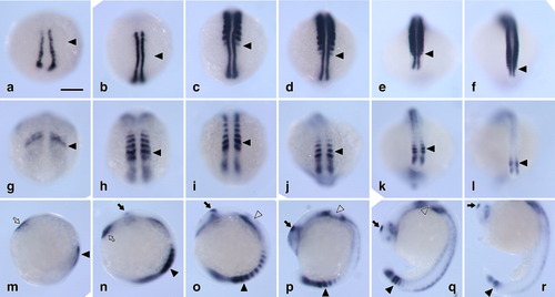
Expression of pcdh10 in zebrafish embryos. Zebrafish embryos were stained by whole-mount in situ hybridization for pcdh10 or myoD. a-f: myoD. g-r: pcdh10. a-l: Dorsal views of the developing somites. m-r: Lateral views. a,g,m, 10 hours postfertilization (hpf); b,h,n, 11.5 hpf; c,i,o, 14 hpf; d,j,p, 16 hpf; e,k,q, 19 hpf; f,l,r, 22 hpf. Closed arrowheads indicate the latest visually segmenting somites. Closed arrows, epiphysis. Open arrowheads, otic vesicles. Open arrows, head mesoderm. Rostral is to the top. Scale bar = 100 μm. |
Expression Data
| Genes: | |
|---|---|
| Fish: | |
| Anatomical Terms: | |
| Stage Range: | 1-4 somites to 26+ somites |
Expression Detail
Antibody Labeling
Phenotype Data
Phenotype Detail
Acknowledgments
This image is the copyrighted work of the attributed author or publisher, and
ZFIN has permission only to display this image to its users.
Additional permissions should be obtained from the applicable author or publisher of the image.
Full text @ Dev. Dyn.