
FIGURE
Fig. 4
- ID
- ZDB-FIG-050121-7
- Publication
- Raymond et al., 2004 - A moving wave patterns the cone photoreceptor mosaic array in the zebrafish retina
- Other Figures
- All Figure Page
- Back to All Figure Page
Fig. 4
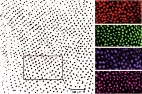
Computational analysis of cone mosaic patterns. (A) Scion Image data file of an adult retinal whole mount processed for in situ hybridization (blue opsin), in which the area selected for analysis is boxed. (B-E) Confocal images of embryonic retinas at ~3dpf processed with red, green, blue or UV opsin probe (pseudo-colored), respectively. |
Expression Data
| Genes: | |
|---|---|
| Fish: | |
| Anatomical Terms: | |
| Stage Range: | Protruding-mouth to Adult |
Expression Detail
Antibody Labeling
Phenotype Data
Phenotype Detail
Acknowledgments
This image is the copyrighted work of the attributed author or publisher, and
ZFIN has permission only to display this image to its users.
Additional permissions should be obtained from the applicable author or publisher of the image.
Full text @ Int. J. Dev. Biol.