
- Title
-
Hox code responsible for the pattering of the anterior vertebrae in zebrafish
- Authors
- Maeno, A., Koita, R., Nakazawa, H., Fujii, R., Yamada, K., Oikawa, S., Tani, T., Ishizaka, M., Satoh, K., Ishizu, A., Sugawara, T., Adachi, U., Kikuchi, M., Iwanami, N., Matsuda, M., Kawamura, A.
- Source
- Full text @ Development
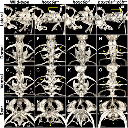
Zebrafish hoxc6a−/−;hoxc6b−/− mutants show severe abnormalities in the third to fourth vertebrae, including the Weberian ossicles. (A-P) The anterior vertebrae were visualized by micro-CT scan in the following group: wild-type (A-D, n=4), hoxc6a−/− (E-H, n=3), hoxc6b−/− (I-L, n=3) and hoxc6a−/−;hoxc6b−/− (M-P, n=4) fish. Adult zebrafish of the same genotype subjected to micro-CT scanning showed essentially similar morphology. Therefore, representative images are shown here. Fish with the desired phenotype were obtained by multiple intercrosses between hoxc6a+/−;hoxc6b+/− fish, and the genotype analysis of the surviving juveniles is shown in Table S1A. The numerical values correspond to the positional order of each vertebra from the first vertebra. In the case of the wild-type zebrafish, unique ossicles attached to each centrum are indicated by a line in A-D. These ossicles attach symmetrically to both sides of the vertebral body. Specifically, the scaphium (sc) is present on the first centrum, and the laterally extending bones known as the lateral process (lp) and intercalarium (ic) are observed on the second centrum. The third centrum possesses a fan-like shaped tripus (tri), and the fourth centrum features a mid-ventrally extending bone known as the os suspensorium (os) and a lateral-ventrally extending bone known as the transverse process of vertebra 4 (tp4). The arrowheads highlight the deformed tripus on the third centrum, which resembles the lateral process on the second centrum in hoxc6a−/− and hoxc6a−/−;hoxc6b−/− mutants. The asterisk indicates the absence of the os suspensorium in hoxc6a−/− and hoxc6a−/−;hoxc6b−/−. The bracket in E,F,M,N indicates the flattened supraneural on the dorsal side of the third centrum in hoxc6a−/− and hoxc6a−/−;hoxc6b−/− mutants. Scale bars: 1 mm. For additional details, micro-CT scan 3D movies are provided in Movies 1,2,3,4. A summary of the phenotypes of these mutants is shown in Table S2. Digital dissection of each vertebra in hoxc6a−/−;hoxc6b−/− is shown in Fig. S3. |

Zebrafish hoxb6a−/−;hoxb6b−/− mutants exhibit shortening of the lateral process in the second vertebra. (A-P) The anterior vertebrae were analyzed by micro-CT scan in wild-type (A-D, n=4), hoxb6a−/− (E-H, n=2), hoxb6b−/− (I-L, n=2) and hoxb6a−/−;hoxb6b−/− (M-P, n=2) fish. Adult zebrafish of the same genotype subjected to micro-CT scanning showed similar morphology; therefore, representative images are shown. Fish were obtained from multiple intercrosses between hoxb6a+/−;hoxb6b+/− fish, and genotype analysis of the surviving juveniles is shown in Table S1B. The numbering indicates the position of each vertebra from the first vertebra. The arrowheads indicate the reduced lateral process on the second centrum in hoxb6a−/− and hoxb6a−/−;hoxb6b−/− mutants. The brackets signify the vertebra with both scaphium and lateral process. Scale bars: 1 mm. For details, micro-CT scan 3D movies are provided in Movies 5,6,7. A summary of the phenotypes of these mutants is shown in Table S2. Digital dissection of each vertebra in hoxb6a−/−;hoxb6b−/− is shown in Fig. S3. ic, intercalarium; lp, lateral process; os, os suspensorium; sc, scaphium; tp4, transverse process of vertebra 4; tri, tripus. |
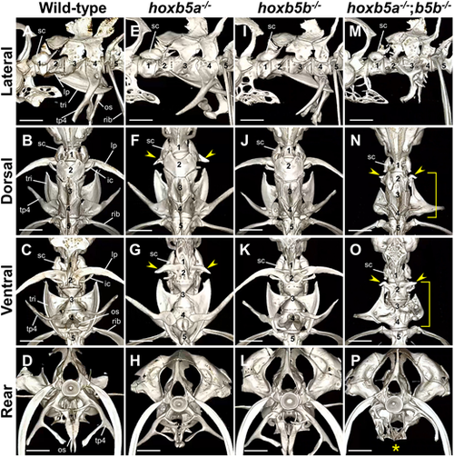
Severe defects in the second to fourth vertebrae of zebrafish hoxb5a−/−;hoxb5b−/−mutants. (A-P) The anterior vertebrae were examined using micro-CT scan in the following groups: wild-type (A-D, n=4), hoxb5a−/− (E-H, n=4), hoxb5b−/− (I-L, n=3) and hoxb5a−/−;hoxb5b−/− (M-P, n=4) adult fish. Representative images are shown. Fish of the desired genotype were obtained by several intercrosses between hoxb5a+/−;hoxb5b+/− fish and the genotype analysis of the surviving juveniles is shown in Table S1C. The numerical values correspond to the positional order of each vertebra from the first vertebra. The arrowheads indicate a significantly shortened lateral process on the second centrum in hoxb5a−/− and hoxb5a−/−;hoxb5b−/− mutants. The brackets indicate malformation of the ossicles on the second to fourth centrum in hoxb5a−/−;hoxb5b−/− mutants. The asterisk indicates the absence of the os suspensorium in hoxb5a−/−;hoxb5b−/− mutants. Scale bars: 1 mm. For additional details, micro-CT scan 3D movies are provided in Movies 8,9,10. A summary of the phenotypes of these mutants is shown in Table S2. Digital dissection of each vertebra in hoxb5a−/−;hoxb5b−/− is shown in Fig. S3. ic, intercalarium; lp, lateral process; os, os suspensorium; sc, scaphium; tp4, transverse process of vertebra 4; tri, tripus |

The absence of the scaphium in the first vertebra in hoxb4a−/−;hoxb5a−/− mutants. (A-H) The anterior vertebrae were examined by micro-CT scan in hoxb4a−/− (A-D, n=4) and hoxb4a−/−;hoxb5a−/− (E-H, n=5) adult fish. Representative images are shown. For hoxb4a−/− and hoxb4a−/−;hoxb5a−/− mutants, fish with the desired genotypes were obtained by multiple intercrosses between each heterozygous fish, and the genotype analysis of the surviving juveniles is shown in Tables S1D,E. The arrowheads indicate the absence of scaphium on the first centrum in hoxb4a−/−;hoxb5a−/−. The asterisk indicates the elongated bone dorsal to the second centrum. Digital dissection of each vertebra in hoxb4a−/−;hoxb5a−/− is shown in Fig. S3. (I,J) Trunk region of wild-type (n=3) and hoxc8a−/− fish (n=5). Adult fish were obtained by several intercrosses between hoxc8a+/− fish, and genotype analysis of the surviving juveniles is shown in Table S1F. The arrowheads indicate the tip of the anterior-most rib. The rib shortening was observed in three of the five hoxc8a mutants. Such rib shortening was not detected in the other Hox mutants isolated in this study. Scale bars: 1 mm in A-H; 2 mm in I,J. For detailed information, micro-CT scan 3D movies are provided in Movies 11,12,13. A summary of the phenotypes of these mutants is shown in Table S2. Digital dissection of each vertebra in hoxb4a−/−;hoxb5a−/− is shown in Fig. S3. ic, intercalarium; lp, lateral process; os, os suspensorium; sc, scaphium; tp4, transverse process of vertebra 4; tri, tripus. |

The second vertebra of medaka hoxc6a−/− mutants shows abnormalities without ribs, similar to the first vertebra. (A-D) Micro-CT scan analysis of the anterior vertebrae in wild-type (n=3) and hoxc6a−/− (n=3) adult medaka. As similar phenotypes were observed for all mutants of the same genotype, a representative individual is shown. The arrowheads indicate the anterior-most pleural ribs attached to vertebrae. In wild-type medaka, the ribs are present from the second vertebra (n=3), but in hoxc6a mutants, the ribs are observed from the third vertebra (n=3). Micro-CT scan 3D movies are provided in Movies 14,15. Scale bars: 1 mm. ep, epipleurals; pp, parapophysis. |

Hox genes responsible for the anterior vertebral identities in mouse, zebrafish and medaka. (A) Diagrams illustrating the typical morphologies of the anterior vertebrae in the zebrafish Hox mutants isolated in this study. The bones that are observed to be abnormal in Hox mutants are highlighted in red, and the missing bones are indicated by dotted lines. Ventral view. (B) Comparisons of Hox genes responsible for the anterior vertebrae in mouse, zebrafish and medaka. Each vertebra is represented by a box, and a triangle attached to the box indicates thoracic vertebrae with ribs. In mice, Hox genes that define the vertebral identities are shown based on malformed vertebrae in Hox knockout mice. In mice, Hox genes from paralog 3 are responsible for vertebral patterning. The specific Hox gene is shown below the vertebrae that are abnormal in individual Hox knockout mice: Hoxb3 (Manley and Capecchi, 1997), Hoxd3 (Condie and Capecchi, 1993), Hoxa4 (Horan et al., 1994; Kostic and Capecchi, 1994), Hoxb4 (Ramirez-Solis et al., 1993), Hoxc4 (Saegusa et al., 1996), Hoxd4 (Horan et al., 1995a), Hoxa5 (Jeannotte et al., 1993), Hoxb5 (Rancourt et al., 1995), Hoxa6 (Kostic and Capecchi, 1994), Hoxb6 (Rancourt et al., 1995) and Hoxc6 (Garcia-Gasca and Spyropoulos, 2000). Multiple paralogous Hox knockout mice showed more severe vertebral abnormalities than single knockout mice. For paralog 3, which has three paralogs, no triple Hox mutants have been reported, but double mutants combining two of the three mutations showed vertebrae with abnormalities (Condie and Capecchi, 1994; Manley and Capecchi, 1997). For paralog 4, triple mutants were reported among the four paralogs, showing C2-C5 vertebrae with abnormalities (Horan et al., 1995b). For paralogs 5 and 6, triple mutants lacking all paralogs have been reported with abnormal vertebrae (McIntyre et al., 2007). In zebrafish, Hox genes that define specific vertebrae, as revealed in this study, are shown below the vertebrae. The frame of the box is black where vertebral abnormalities were observed in double Hox mutants. In medaka, hoxc6a is responsible for the second vertebra. |