
|
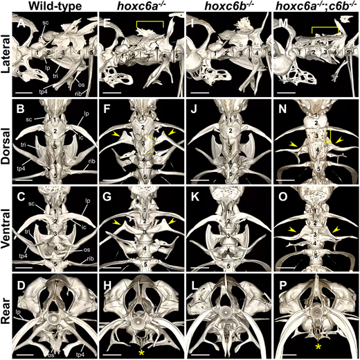
Fig. 1 Zebrafish hoxc6a−/−;hoxc6b−/− mutants show severe abnormalities in the third to fourth vertebrae, including the Weberian ossicles. (A-P) The anterior vertebrae were visualized by micro-CT scan in the following group: wild-type (A-D, n=4), hoxc6a−/− (E-H, n=3), hoxc6b−/− (I-L, n=3) and hoxc6a−/−;hoxc6b−/− (M-P, n=4) fish. Adult zebrafish of the same genotype subjected to micro-CT scanning showed essentially similar morphology. Therefore, representative images are shown here. Fish with the desired phenotype were obtained by multiple intercrosses between hoxc6a+/−;hoxc6b+/− fish, and the genotype analysis of the surviving juveniles is shown in Table S1A. The numerical values correspond to the positional order of each vertebra from the first vertebra. In the case of the wild-type zebrafish, unique ossicles attached to each centrum are indicated by a line in A-D. These ossicles attach symmetrically to both sides of the vertebral body. Specifically, the scaphium (sc) is present on the first centrum, and the laterally extending bones known as the lateral process (lp) and intercalarium (ic) are observed on the second centrum. The third centrum possesses a fan-like shaped tripus (tri), and the fourth centrum features a mid-ventrally extending bone known as the os suspensorium (os) and a lateral-ventrally extending bone known as the transverse process of vertebra 4 (tp4). The arrowheads highlight the deformed tripus on the third centrum, which resembles the lateral process on the second centrum in hoxc6a−/− and hoxc6a−/−;hoxc6b−/− mutants. The asterisk indicates the absence of the os suspensorium in hoxc6a−/− and hoxc6a−/−;hoxc6b−/−. The bracket in E,F,M,N indicates the flattened supraneural on the dorsal side of the third centrum in hoxc6a−/− and hoxc6a−/−;hoxc6b−/− mutants. Scale bars: 1 mm. For additional details, micro-CT scan 3D movies are provided in Movies 1,2,3,4. A summary of the phenotypes of these mutants is shown in Table S2. Digital dissection of each vertebra in hoxc6a−/−;hoxc6b−/− is shown in Fig. S3.