FIGURE SUMMARY
Title

Identification, Expression and Evolutional Analysis of Two cyp19-like Genes in Amphioxus

Authors
Wang, Y., Lin, J., Li, W., Ji, G., Liu, Z.
Source
Full text @ Animals (Basel)

Complete ORF nucleotide sequences of two cyp19-like fragments in amphioxus. (A): The cyp19-like1 ORF in amphioxus, with a size of 1491bp. (B): The cyp19-like2 ORF in amphioxus, with a size of 1482bp.

Comparison of the gene structures of cyp19a1a, cyp19a1b, and aromatase-like genes in zebrafish and Florida amphioxus.

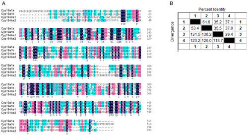
Alignment of amino acid sequences of cyp19 in zebrafish and amphioxus. (A): Amino acid sequence alignment; (B): sequence similarity comparison. 1 represents cyp19a1a, 2 represents cyp19a1b, 3 represents cyp19-like1, and 4 represents cyp19-like2.

Prediction of Cyp19 structures in zebrafish and amphioxus. (A): SMART prediction of domains in zebrafish Cyp19a1a (a), Cyp19a1b (b), and amphioxus Cyp19-like1 (c), Cyp19-like2 (d); (B): PHYRE2 prediction of three-dimensional structures in zebrafish Cyp19a1a (a), Cyp19a1b (b), and amphioxus Cyp19-like1 (c), Cyp19-like2 (d).

Analysis of synteny and phylogeny of Cyp superfamily across different species. (A): Location maps of Cyp19 on amphioxus and other vertebrate chromosomes. Boxes represent genes, and the direction of the boxes indicates the orientation of the genes. (B): Phylogenetic analysis of Cyp superfamily across different species. Sequence sources: see Supplemental Table S1.

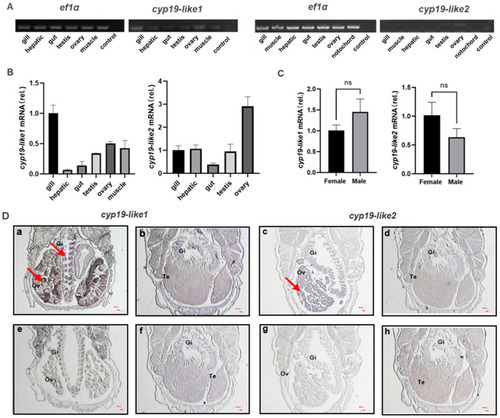
Tissue expression and localization of cyp19-like in amphioxus. (A): Semi-quantitative PCR detection of cyp19-like gene expression in different tissues of amphioxus. (B): Real-time PCR detection of cyp19-like gene expression in different tissues of amphioxus, including gill, hepatic, gut, testis, ovary, muscle, and notochord; (C): expression of cyp19-like in the gills of male and female amphioxus. ns indicates no significant difference. (D): ISH results of cyp19-like on paraffin sections of amphioxus. (ad) show antisense probe hybridization results; (eh) show sense probe hybridization results (negative control). Gi: gill; Ov: ovary; Te: testis. Arrows indicate positive signals. Scale bar: 100 μm.

Expression of cyp19-like in amphioxus at different developmental stages. (A): Tissue morphology and HE staining results at different stages of gonad development in amphioxus. Gi: gill; Go: gonad; arrows indicate gonadal primordium. (B): Real-time PCR detection of cyp19-like expression at different stages of gonad development in amphioxus. T1, T2, and T3 represent the primordial, developing, and mature gonad stages, respectively.

Antibody preparation and protein expression of amphioxus Cyp19-like. (A): Designated sites for immunogenic peptides which are indicated with arrows. (B): Western blot results of Cyp19-like in amphioxus. (C): Immunohistochemical results of Cyp19-like in amphioxus. T1, T2, and T3 represent the primordial, developing, and mature gonad stages, respectively. a–f show Cyp19-like1 immunohistochemical results, while g–l show Cyp19-like2 immunohistochemical results. a–c, g–i are experimental groups, while d–f, j–l are control groups. Gi: gill; Go: gonad; arrows indicate positive signals. Scale bar: 100 μm. (D): Subcellular localization of amphioxus Cyp19-like in HEK cells using LSCM. The expression of Cyp19-like is depicted in green; nuclei were counterstained with DAPI (blue). Red arrow indicating positive signal. Scale bar: 10 μm.

Acknowledgments
This image is the copyrighted work of the attributed author or publisher, and ZFIN has permission only to display this image to its users. Additional permissions should be obtained from the applicable author or publisher of the image. Full text @ Animals (Basel)