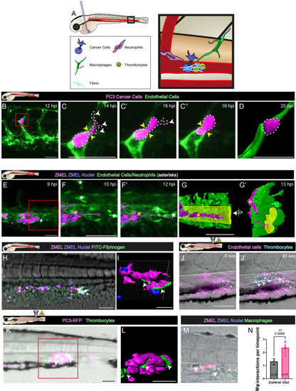
CLEM views of cancer cells interacting closely with host innate immune cells and vasculature of the pre-metastatic niche. (A) View of human prostate cancer cells (magenta) post injection into the vasculature (green) of a 3 dpf larval zebrafish. The box indicates the higher-magnification image shown in B. (B) Overlay of light microscopy and transmission electron microscopy (TEM) images of this region. The red box in B indicates the higher-magnification image shown in C. (C,C′) Direct interactions between a macrophage (Mϕ, cyan) and a cancer cell (PC3, magenta), including interdigitation (arrowheads, C′) between the membranes of these cells. RBC, red blood cell. (D) TEM image of a human cancer cell (PC3, magenta) interacting closely with a zebrafish thrombocyte (T, green). (D′) Higher-magnification image of the same cancer cell shown in D, 5 µm deeper. The inset shows close membrane contacts with possible membrane exchange (arrowheads). (E,E′) Schematic of cancer cell integration into the larval vasculature. (F,F′) Fluorescence (F) and brightfield (F′) still images from a time-lapse movie of a human cancer cell (magenta) integrating into a vessel (green) (Movie 4). The dashed line indicates the cancer lumen. (G–G″) Multi- and split-channel still images from the same time-lapse. (H) Imaris rendering of the same cancer cell undergoing vascular integration. Arrowheads in G,G′,H indicate cancer cell extensions into the vasculature. White asterisks in F–H indicate vessel-associated macrophages, whereas yellow asterisks indicate blood cells within the lumen. Images are representative of ≥3 independent experiments (A–D′), and a single experiment (F–H). Scale bars: 500 µm (A); 10 µm (C,C′); 2.5 µm (C′ inset); 5 µm (D,D′); 50 µm (F–H).
|