IMAGE

Fig. 2.

ID
ZDB-IMAGE-231010-8
Source
Figures for Ward et al., 2023
Image
Figure Caption

Fig. 2.

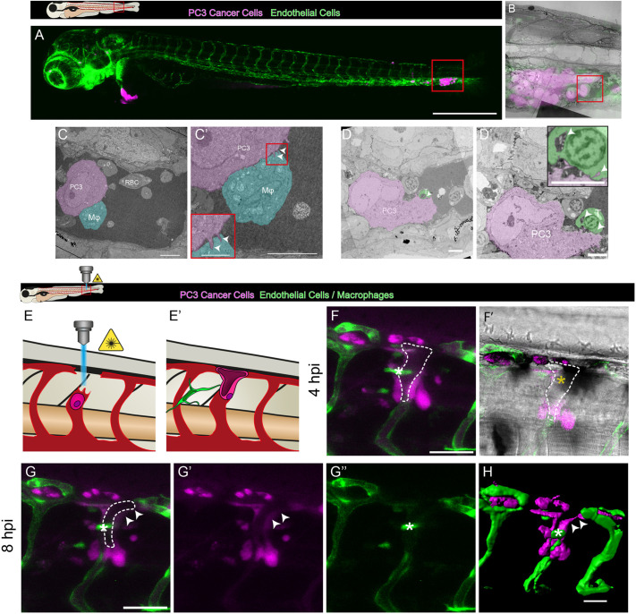
CLEM views of cancer cells interacting closely with host innate immune cells and vasculature of the pre-metastatic niche. (A) View of human prostate cancer cells (magenta) post injection into the vasculature (green) of a 3 dpf larval zebrafish. The box indicates the higher-magnification image shown in B. (B) Overlay of light microscopy and transmission electron microscopy (TEM) images of this region. The red box in B indicates the higher-magnification image shown in C. (C,C′) Direct interactions between a macrophage (Mϕ, cyan) and a cancer cell (PC3, magenta), including interdigitation (arrowheads, C′) between the membranes of these cells. RBC, red blood cell. (D) TEM image of a human cancer cell (PC3, magenta) interacting closely with a zebrafish thrombocyte (T, green). (D′) Higher-magnification image of the same cancer cell shown in D, 5 µm deeper. The inset shows close membrane contacts with possible membrane exchange (arrowheads). (E,E′) Schematic of cancer cell integration into the larval vasculature. (F,F′) Fluorescence (F) and brightfield (F′) still images from a time-lapse movie of a human cancer cell (magenta) integrating into a vessel (green) (Movie 4). The dashed line indicates the cancer lumen. (G–G″) Multi- and split-channel still images from the same time-lapse. (H) Imaris rendering of the same cancer cell undergoing vascular integration. Arrowheads in G,G′,H indicate cancer cell extensions into the vasculature. White asterisks in F–H indicate vessel-associated macrophages, whereas yellow asterisks indicate blood cells within the lumen. Images are representative of ≥3 independent experiments (A–D′), and a single experiment (F–H). Scale bars: 500 µm (A); 10 µm (C,C′); 2.5 µm (C′ inset); 5 µm (D,D′); 50 µm (F–H).

Acknowledgments
This image is the copyrighted work of the attributed author or publisher, and ZFIN has permission only to display this image to its users. Additional permissions should be obtained from the applicable author or publisher of the image. Full text @ J. Cell Sci.