FIGURE SUMMARY
Title

Identification of chicken LOC420478 as Bucky ball equivalent and potential germ plasm organizer in birds

Authors
Klein, S., Dosch, R., Altgilbers, S., Kues, W.A.
Source
Full text @ Sci. Rep.

Regions of partial amino acid homology for chicken and zebrafish Bucky ball. (a) Homology regions between the predicted chicken (upper panel) and zebrafish Bucky ball amino acid sequences (lower panel). (b) Amino acid sequence of LOC 420748 with marked strings of homology to the zebrafish Bucky ball sequence and with highlighted positions of the predicted nuclear localization sequences. The nuclear localization sequences were predicted by cNLS mapper (https://nls-mapper.iab.keio.ac.jp/cgi-bin/NLS_Mapper_form.cg).

High resolution confocal imaging of single channels for the reporter fluorescence (green fluorescence proteins, green label), for immunocytochemical Bucky ball reactivity in DF1 cells after transfection with different fusion plasmids (red label) and nuclear label using SIR (cyan label): (A) two examples for the GFP only plasmid; (B) two examples for the chicken Bucky ball—venus fusion plasmid; (C) two examples for the zebrafish Bucky ball- GFP fusion plasmid; D negative control sample of chicken Bucky ball transfected cells incubated without primary Bucky ball antibody; Left larger frames represents the overlay of all three channels (1,4), right upper frames represents the overlay of the GFP reporter and Bucky ball channel (2,5), right lower frames (3,6) represent the overlay of the nuclear marker and Bucky ball label). (E,F) Examples of DF1 cells transfected with chicken Bucky ball plasmid without Venus reporter; (G,H) examples of DF1 cells transfected with zebrafish Bucky ball without GFP reporter, overlay of all three channels each; all images were taken with a 63× oil immersion 1.6 NA objective.

Expression of Bucky ball after transfection of fusion plasmids in vitro and in embryonic stages around oviposition in vivo (a) Bucky ball fusion-constructs form insoluble fusion proteins. Bucky ball fusion construct and controls (EGFP/Venus only) were transfected in DF1 cells, respectively. After 5 days, the cells were harvested, and total extracts, insoluble, and soluble fractions were assessed by Western blotting with an anti-GFP antibody. Controls (EGFP/Venus only, lanes 1, 4, and 7), zebrafish Bucky ball-EGFP (lanes 2, 5, and 8), and chicken Bucky ball-Venus fusion constructs. M, size marker with visible bands at 20, 40, 50, 60, 100, 150 kDalton. Note, the Bucky ball fusion proteins appear in the insoluble fraction (lanes 4, and 5) and migrate much slower than expected for the calculated molecular weights of 101 kDa (zBucky ball-EGFP) and 116 kDa (cBucky ball-Venus). The EGFP/Venus control shows a protein at the expected molecular weight of 27 kDa. (b) Detection of embryonically transcribed Bucky ball in the chicken blastoderm. The upper panel refers to Bucky ball and the lower panel to GAPDH transcripts. Samples 1 to 3 were generated from stage EGK X28 laid egg blastoderms, and samples 4 to 6 from 24 h incubated embryos (stage HH 627). DNA-ladder in lanes (M): NEB 100 bp ladder N3231, (B) negative control amplification without template.

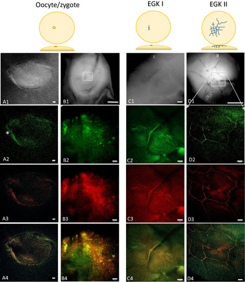
Accumulation of Bucky ball protein from the oocyte to early cleavage stages (EGK I/II) in chicken embryos. The upper panel represents differential interference (A1) and bright field images (B1,C1,D1) of the germinal vesicle (A1), the total fragment containing the germinal disc (B1) and the germinal disc regions (C1,D1). Frames indicate the region of the higher magnified fluorescent images in the lower panels. Freshly ovulated oocytes are characterized by a diffuse distribution of the Bucky ball protein from a condensed outline of the germinal vesicle (A2). At the outline, a higher Bucky ball concentration at one side (*) is apparent. After zygote formation, when the fertilized oocyte reached the isthmus part of the oviduct 2 to 2.5 h after fertilization, Bucky ball protein concentrates in granular patches in the center of the germinal disc where first cleavage furrows will be formed (B2). The first cleavage furrow is lined by tightly aggregated Bucky ball, and in perpendicular orientation, the Bucky ball granules demarcate the predicted next furrow (C2). During the next furrow divisions of stage I and II, further Bucky ball protein condenses in the cleavage furrows in the center of the forming embryo (D2). There is no clear demarcation of cellular outlines yet. This pattern of protein distribution is highly colocalizing to CVH (A3–C3), one of the first known germ plasm proteins as visualized in the overlay in A4–C4). Scales represent for (A) = 20 µm, (B1) = 200 µm; (C) = 50 µm; (D1) = 200 µm, the panels (B2–B4) = 20 µm, and (D2–D4) = 50 µm.

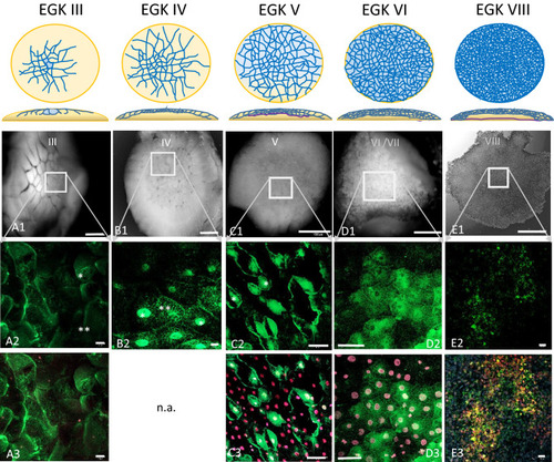
Dynamic localization of Bucky ball around major zygotic genome activation. The upper panel represents schemes for the formation of cells from stage EGK III, where first centrally located cells get separated to stage VIII, the second stage of area pellucida formation phase as described by Eyal-Giladi and Kochav28,29 (A1) An eccentrical position (rectangular frame in (A1), upper left panel) of the cleavage center within the chicken germinal disc region. With the ongoing cell divisions during EGK III, Bucky ball protein granules start to accumulate in the cytoplasm of the medial cleavage center in first laterally and ventrally closed cells (* in A2). The protein is not found accumulating around or in the nuclei stained with SIR and visualized in red. In the more lateral cells, the protein is still located in or around the cleavage furrows (**). At seven to eight hours after fertilization (hpf) during stage EGK IV, Bucky ball is first observed in the nuclei of central cells with very high intensity of labeling (B2*), whereas at the surrounding cells Bucky ball is still located in the cytoplasm (B2**). The same pattern of intracellular protein localization of Bucky ball was recorded in stage V embryos (C1 overview, C2 magnified central frame with nuclear and cytoplasmic labeling of Bucky ball. (A3,C3–E3) demonstrate SIR counterstained nuclei and the concentration of Bucky ball in and around them. After around 12 hpf, when embryos developed to stage VI, Bucky ball seems to decrease in intensity overall and the nuclear signals disappear (D2). This process of signal reduction proceeds further to stage VIII and no nuclear labeling remained (E2). The basal membrane is separating the yolk from the developing embryo, and starts growing from the middle to the edges of the disc at stage V, (purple line, scheme above column C) and reaches the outline of the disc at stage VI (scheme, column D). The subgerminal cavity is formed between the embryo and the basal membrane via cell shedding to the outline forming the area pellucida from stage VII onwards (scheme, column (E)). Scales (A1–E1) = 1 mm, (A2,A3,C2,C3) = 50 µm; (B2,D2,D3) = 10 µm, (E2,E3) = 20 µm.

Co-localization of Bucky ball and CVH during stages EK I to EK III. During the first two cleavage stages (EK I and EK III), Bucky ball strictly co-localized to CVH within the cleavage furrows and in condensed granules in the cleavage center A: (A1) stitched fluorescent overlay images of the central region of an EGK II embryo with the indicated region of high resolution images shown in (A2–A4) (A2) Bucky ball, (A3) CVH, (A4) overlay. (A2—A4) are included in a preprint51. During the next stage, EK III, both germ plasm proteins translocate into the cytoplasm of the first forming central cells B: (B1) transmission image of the total embryonic disc at stage EGK III with indicated central region for high resolution fluorescent imaging: (B2) Bucky ball, (B3) CVH, (B4) overlay. Asterisk indicate the diffusion of Bucky ball and CVH proteins into the cytoplasm. Scales 20 µm, except (B1) = 500 µm.

Overview of an intrauterine embryo (EGK III) with two frames of the central region with Bucky ball immunolabeling in contrast to severely reduced immuno-fluorescence at the lateral outline of furrows. The two frames in the central region show the cells with Bucky ball immunofluorescence in cytoplasmic granular distribution. The lateral frame, documented at the exactly comparable acquisition conditions, does not provide signals above background. Scale = 500 µm.

Co-localized Bucky ball and CVH expression in stage EGK IV. This composition of eight stitched images visualizes in the magnified center of a stage EGK IV embryo (frame of the overview in (A) the concentrated nuclear localization of Bucky ball (B) and CVH (C) within the central cells of the germinal disc surrounded by cells with cytoplasmic germ plasm of Bucky ball and CVH. In (D) the overlay with clear co-localization of Bucky ball (E) and CVH (F) in cells of the nuclear region of EGK IV embryos is shown. Numbers with nuclear region labels exceed 20 cells repeatedly. Scale in (A): 500 µm, scales (D–F): 20 µm.

Bucky ball and CVH are no longer co-localized within the nuclear region of primordial germ cells during the area pellucida formation. 7A, in EGK VII embryos, at the beginning of area pellucida formation, Bucky ball (green) in the center of the germinal disc diffused back into the cytoplasm (A2), while CVH (red, A4) is contained predominantly in the nuclei. This is visualized clearly in the overlay (A3) as a red center of the germline predisposed cells surrounded by the green Bucky ball fluorescence. Blue frames with asterisk indicate only CVH labeled without Bucky ball at the lateral edges of the central region. DAPI as nuclear marker visualizes the central concentration of the germline cells and as white overlay the nuclear localization of CVH and DAPI in the germline cells. This pattern is stable until oviposition at stage X (B). Scales in (A1) = 200 µm, (B1): 500 µm, (A2–A4) 20 µm, (B2–B4): 100 µm.

Acknowledgments
This image is the copyrighted work of the attributed author or publisher, and ZFIN has permission only to display this image to its users. Additional permissions should be obtained from the applicable author or publisher of the image. Full text @ Sci. Rep.