IMAGE

Figure 5

ID
ZDB-IMAGE-221018-38
Source
Figures for Klein et al., 2022
Image
Figure Caption

Figure 5

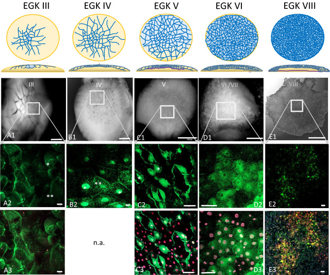
Dynamic localization of Bucky ball around major zygotic genome activation. The upper panel represents schemes for the formation of cells from stage EGK III, where first centrally located cells get separated to stage VIII, the second stage of area pellucida formation phase as described by Eyal-Giladi and Kochav28,29 (A1) An eccentrical position (rectangular frame in (A1), upper left panel) of the cleavage center within the chicken germinal disc region. With the ongoing cell divisions during EGK III, Bucky ball protein granules start to accumulate in the cytoplasm of the medial cleavage center in first laterally and ventrally closed cells (* in A2). The protein is not found accumulating around or in the nuclei stained with SIR and visualized in red. In the more lateral cells, the protein is still located in or around the cleavage furrows (**). At seven to eight hours after fertilization (hpf) during stage EGK IV, Bucky ball is first observed in the nuclei of central cells with very high intensity of labeling (B2*), whereas at the surrounding cells Bucky ball is still located in the cytoplasm (B2**). The same pattern of intracellular protein localization of Bucky ball was recorded in stage V embryos (C1 overview, C2 magnified central frame with nuclear and cytoplasmic labeling of Bucky ball. (A3,C3–E3) demonstrate SIR counterstained nuclei and the concentration of Bucky ball in and around them. After around 12 hpf, when embryos developed to stage VI, Bucky ball seems to decrease in intensity overall and the nuclear signals disappear (D2). This process of signal reduction proceeds further to stage VIII and no nuclear labeling remained (E2). The basal membrane is separating the yolk from the developing embryo, and starts growing from the middle to the edges of the disc at stage V, (purple line, scheme above column C) and reaches the outline of the disc at stage VI (scheme, column D). The subgerminal cavity is formed between the embryo and the basal membrane via cell shedding to the outline forming the area pellucida from stage VII onwards (scheme, column (E)). Scales (A1–E1) = 1 mm, (A2,A3,C2,C3) = 50 µm; (B2,D2,D3) = 10 µm, (E2,E3) = 20 µm.

Acknowledgments
This image is the copyrighted work of the attributed author or publisher, and ZFIN has permission only to display this image to its users. Additional permissions should be obtained from the applicable author or publisher of the image. Full text @ Sci. Rep.