
- Title
-
Granulocyte Colony-Stimulating Factor Mediated Regulation of Early Myeloid Cells in Zebrafish
- Authors
- Meier, A.B., Basheer, F., Sertori, R., Laird, M., Liongue, C., Ward, A.C.
- Source
- Full text @ Front Biosci (Landmark Ed)

Alternative G-CSFR pathways in myelopoiesis. (A–D) Fluorescent microscopy images of representative 22 hpf Tg (mpo::GFP) embryos injected with either scr Mo (A), csf3a Mo (B), csf3b Mo (C) or csf3r Mo (D). (E,G) Quantitation of the number of mpo+ cells in embryos injected with morpholinos (E) or treated with specific inhibitors (G). (F,H) Quantitation of relative migration of mpo+ cells in embryos injected with morpholinos (F) or specific inhibitors (H). (I–L) Fluorescent microscopy images of representative 48 hpf Tg (lyz::DsRed) embryos injected with either scr Mo (I), csf3a Mo (J), csf3b Mo (K) or csf3r Mo (L). (M,N) Quantitation of lyz+ cells in embryos injected with morpholinos (M) or specific inhibitors (N). In panels E–H and M–N, results are shown for individual embryos along with mean and SEM in red, with the level of statistical significance relative to controls indicated (*: p |

Role of alternative G-CSFR ligands in response to wounding. (A–L) Fluorescent microscopy images of representative Tg (mpo::GFP) embryos injected with either scr Mo (A–C), csf3a Mo (D–F), csf3b (G–I) or csf3r Mo (J–L) at the indicated times post wounding. (M,N) Quantitation of recruitment of mpo+ (M) and lyz+ (N) cells to the wounding site at 0, 2, 4, 6 and 8 hours post wounding (hpw) in Tg (mpo::GFP) and Tg (lyz::DsRed) embryos, respectively, that had been injected with the indicated morpholinos, showing mean and S.E.M. along with statistical significance of gene-specific in comparison to scr Mo (*: p |

Role of alternative G-CSFR pathways in response to wounding. (A–I) Fluorescent microscopy images of representative Tg (mpo::GFP) embryos pre-treated with either DMSO (A–C), AG490 (D–F) or LY294002 (G–I) at the indicated times post wounding. (J,K) Quantitation of recruitment of mpo+ (J) and lyz+ (K) cells to the wounding site at 0, 2, 4, 6 and 8 hours post wounding (hpw) in Tg (mpo::GFP) and Tg (lyz::DsRed) embryos, respectively, that had been pre-treated with the indicated inhibitors, showing mean and S.E.M. along with statistical significance of inhibitor in comparison to DMSO control (*: p |
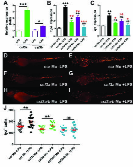
Role of alternative G-CSFR ligands in emergency myelopoiesis. (A–C) qRT |

Role of alternative G-CSFR pathways in emergency myelopoiesis. (A–H) Fluorescent microscopy images of representative Tg (mpo::GFP) embryos treated with either DMSO (A,B), AG490 (C,D), LY294002 (E,F) or PP2 (G,H) either without (-) (A,C,E,G) or with (+) (B,D,F,H) LPS treatment. (I,J) Quantitation of mpo+ cells in Tg (mpo::GFP) embryos (I) or lyz+ cells in Tg (lyz::DsRed) embryos (J) treated with the indicated agents either without (-) with (+) LPS treatment, showing individual embryos as well as mean and S.E.M. along with statistical significance (*: p |