FIGURE SUMMARY
Title

Bloom syndrome helicase contributes to germ line development and longevity in zebrafish

Authors
Annus, T., Müller, D., Jezsó, B., Ullaga, G., Németh, B., Harami, G.M., Orbán, L., Kovács, M., Varga, M.
Source
Full text @ Cell Death Dis.

RecQ homologs in the zebrafish genome and their expression.

A Phylogenetic relationships of the five RecQ homologs that can be identified in the zebrafish genome based on similarities between the helicase ATPase and helicase C-terminal domains. (M. hungatei Hel308 DNA helicase was used as an external reference for eukaryotic RecQs). B, C The expression of genes related to double-strand DNA breaks during zebrafish development. Fragments per kilobase of exon model per million reads mapped (FPKM) and transcripts per kilobase million (TPM) values of the different genes at given stages as shown by the datasets in the respective papers (White et al., 2017; Winata et al., 2018). D Spatial distribution of blm RNA during early stages of development detected by whole-mount in situ hybridization. The number of embryos showing the expression pattern is indicated in the upper right corner. (All lateral views; for shield and 16 somite stages embryos ventral is to the left and dorsal to the right, while for 1 and 2 dpf embryos anterior is to the left and posterior is to the right, respectively. Scale bars: 150 μm.). E Schematic domain structure of zebrafish Blm and the position of the p.Thr101Leufs*4 mutation. (RQC RecQ-C-terminal domain, HRDC helicase and RNaseD C-terminal.). F Sanger sequencing shows the presence of the c.301_304delACAAinsTT allele (elu15) in exon 4 of a blm/− embryo.

EXPRESSION / LABELING:
Gene:
Fish:
Anatomical Terms:
Stage Range: 14-19 somites to Prim-5

Loss of Blm function does not increase sensitivity to DNA damage, however severely reduces lifespan in zebrafish.

A, A′ Anti-γ-H2AX labeling in the head and trunk of 3 dpf untreated wild-type (A) and blm−/ embryos (A′) (Scale bar: 500 μm). B, B′ Anti-γ-H2AX labeling in the head and trunk of 3 dpf wild-type (B) and blm−/− (B′) animals after treatment with the DNA interstrand cross-linking agent diepoxybutane (DEB) (Scale bar: 500 μm). C, D γ-H2AX-positive foci in the head and trunk of different blm genotypes under control circumstances (C) and after DEB treatment (D) (ns not significant). E Phenotypes observed after gamma irradiation: Class 1—wild-type; Class 2—mild necrosis in the tectum and smaller eyes; Class 3—heavy necrosis all over the body, heart edema. Untreated controls were all Class 1. F, G The distribution of phenotype severity following gamma irradiation (F) and DEB treatment (G) (ns not significant). H Survival probability graphs of different blm genotypes. (Pairwise p values were calculated with the log-rank test, using Benjamini & Hochberg adjustment. dpf days post-fertilization).

Homozygous <italic>blm</italic> mutants develop into males with fertility defects.

A The sex ratio of offsprings derived from blm+/ incrosses suggests a role for Blm in zebrafish sex determination and/or gonad differentiation. Data from two incrosses combined is shown. (For individual incrosses, see Supp Fig. 2A). B Complete lack of females among blm−/ mutant fish in the absence of a functional Tp53 indicates that the effect of Blm is Tp53-independent. (Pairwise p values were calculated with Chi-square test; ns not significant. Data from two incrosses combined are shown. For individual incrosses, see Supplementary Fig. 2B). C The number of GFP-positive germ cells (GCs) in the gonad of blm+/+ and blm+/− individuals was substantially higher than that of their blm−/− siblings. All individuals were on a Tg(ddx4:egfp) background at the age of 1-month post-fertilization (scale bar: 100 μm). D Gonadal GC counts of different blm genotypes as determined on a Tg(ddx4:egfp) background at the size of 5–6 mm. While GC counts of heterozygous blm+/ individuals were similar to those of wild types, homozygous blm−/− individuals showed significantly lower numbers (p = 2.5e-03; Welch t-test). E The ratio of fertilized embryos in a total of 51 crosses using males of different blm genotypes. F, G Toluidine Blue staining of testis sections from wild-type (F) and blm/ mutant (G) fish. Asterisks denote spermatozoa clusters of normal densities. Homozygous mutant blm/− testes showed a drastic reduction of mature spermatozoa (scale bar: 50 μm). Labels: Fe female, M male, GC germ cell.

Blm loss-of-function results in meiotic defects during spermatogenesis in zebrafish males.

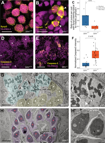
A, B Representative Sycp3 immunostainings of wild-type and blm−/ spermatogonial cysts. In wild-type cysts (A) patterns typical for meiotic prophase I can be detected, whereas in mutant cysts (B) aberrant Sycp3 patterns (white arrowheads) can be observed (n = 8 different wild-type and blm−/− samples were tested each). TO-PRO-3 staining (red) denotes nuclei (scale bar: 8 μm). C Ratio of nuclei showing Sycp3 immunostaining patterns characteristic for pachytene. A comparison of wild-type and mutant testes suggests that blm/ are defective in entering pachytene. D, E Cells undergoing programmed cell death as shown by Caspase-3 (green) staining in wild-type (D) and blm−/− testes (E). TO-PRO-3 staining (red) denotes nuclei (n = 6 different wild-type and blm−/ testis lobes were tested each). Asterisks denote spermatozoa clusters (Scale bar: 50 μm). F Normalized Caspase-3 stainings of wild-type and mutant testes suggest increased apoptosis in the absence of Blm. G, G′ Electron microscopic images of representative spermatogenic cysts with spermatocytes in meiotic prometaphase (cyan) and other early stages of meiosis I (yellow) from wild-type males. White asterisks denote condensed chromosomal structures, white hash symbols indicate nuclei in early meiotic prophase I (see Zhang et al., 2014). The white arrowhead in G′ indicates a cytoplasmic bridge between spermatocytes (scale bar: 5 μm). H, H′ Electron micrographs of spermatogenic cysts of blm−/ mutant males with spermatocytes (purple) showing aberrantly condensed chromatin (red asterisk), which was found to be the characteristic feature of mutant testes. Note that there are no nuclear envelopes observed around chromatin condensates, indicating that this phenomenon is related to cell division (see also Supplementary Fig. 5) (scale bar: 5 μm).

A model for the multiple roles of Blm during zebrafish gonadal development.

During the early stages of ontogenesis ddx4 (formerly vasa) expressing primordial germ cells (PGC) proliferate and migrate from their initial positions adjacent to the yolk syncytial layer to the gonadal mesoderm. There their proliferation continues, while a varying number of these GCs differentiate into stage 1a oocytes resulting in the development of the “juvenile ovary”. A In wild-type fish the number of GCs may further increase to the point where it crosses the threshold required for female fate-determining pathways to activate, resulting in the development of mature ovaries with GC-derived ovarian stem cells (OSC) and mature oocytes. If the threshold is not reached either due to insufficient GC proliferation, or none at all, a major shift in the gonadal environment occurs through which oocytes (and possibly even a subset of GCs) are eliminated via apoptosis. Subsequently, the formation of spermatogonial stem cells (SSC) and Sertoli cells occurs either by (i) differentiation of common OSC and SSC precursors (gonadal stem cells) into SSCs that in turn induce Sertoli cell differentiation in the soma; or (ii) by the transdifferentiation of somatic follicular cells into Sertoli cells that promote SSC differentiation and expansion from gonadal stem cells, thus leading to testis development. A’ In the wild-type testis, SSCs are enveloped by Sertoli cells where they further differentiate into spermatogonia. In this structure, known as a cyst, spermatogonia go through mitotic expansion followed by synchronous meiosis into spermatids. B In Blm loss-of-function individuals, the complete lack of females might be caused by two major factors: (i) the number of GCs never reaches the threshold necessary for female development; and/or (ii) meiotic processes necessary for the formation of oocytes might be compromised in absence of Blm. As we do not know whether the gonads of blm−/− zebrafish do make the initial “detour” through the juvenile ovary stage or not, both possibilities are indicated. B′ A potential explanation for the subfertility of blm−/− males is that in the absence of Blm accurate DNA repair during spermatogonial meiosis is hindered, and therefore cell death is induced.

Acknowledgments
This image is the copyrighted work of the attributed author or publisher, and ZFIN has permission only to display this image to its users. Additional permissions should be obtained from the applicable author or publisher of the image. Full text @ Cell Death Dis.