IMAGE

Fig. 3

ID
ZDB-IMAGE-220430-7
Genes
Source
Figures for Annus et al., 2022
Image
Figure Caption

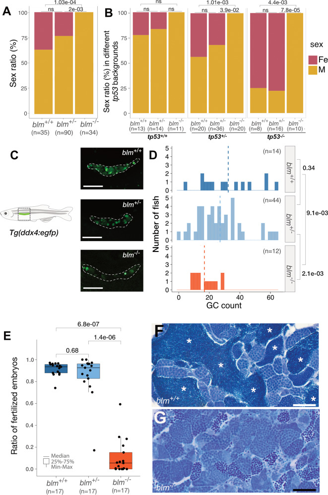
Fig. 3 Homozygous <italic>blm</italic> mutants develop into males with fertility defects.

A The sex ratio of offsprings derived from blm+/ incrosses suggests a role for Blm in zebrafish sex determination and/or gonad differentiation. Data from two incrosses combined is shown. (For individual incrosses, see Supp Fig. 2A). B Complete lack of females among blm−/ mutant fish in the absence of a functional Tp53 indicates that the effect of Blm is Tp53-independent. (Pairwise p values were calculated with Chi-square test; ns not significant. Data from two incrosses combined are shown. For individual incrosses, see Supplementary Fig. 2B). C The number of GFP-positive germ cells (GCs) in the gonad of blm+/+ and blm+/− individuals was substantially higher than that of their blm−/− siblings. All individuals were on a Tg(ddx4:egfp) background at the age of 1-month post-fertilization (scale bar: 100 μm). D Gonadal GC counts of different blm genotypes as determined on a Tg(ddx4:egfp) background at the size of 5–6 mm. While GC counts of heterozygous blm+/ individuals were similar to those of wild types, homozygous blm−/− individuals showed significantly lower numbers (p = 2.5e-03; Welch t-test). E The ratio of fertilized embryos in a total of 51 crosses using males of different blm genotypes. F, G Toluidine Blue staining of testis sections from wild-type (F) and blm/ mutant (G) fish. Asterisks denote spermatozoa clusters of normal densities. Homozygous mutant blm/− testes showed a drastic reduction of mature spermatozoa (scale bar: 50 μm). Labels: Fe female, M male, GC germ cell.

Figure Data
Acknowledgments
This image is the copyrighted work of the attributed author or publisher, and ZFIN has permission only to display this image to its users. Additional permissions should be obtained from the applicable author or publisher of the image. Full text @ Cell Death Dis.