FIGURE SUMMARY
Title

The endosomal RIN2/Rab5C machinery prevents VEGFR2 degradation to control gene expression and tip cell identity during angiogenesis

Authors
Kempers, L., Wakayama, Y., van der Bijl, I., Furumaya, C., De Cuyper, I.M., Jongejan, A., Kat, M., van Stalborch, A.D., van Boxtel, A.L., Hubert, M., Geerts, D., van Buul, J.D., de Korte, D., Herzog, W., Margadant, C.
Source
Full text @ Angiogenesis

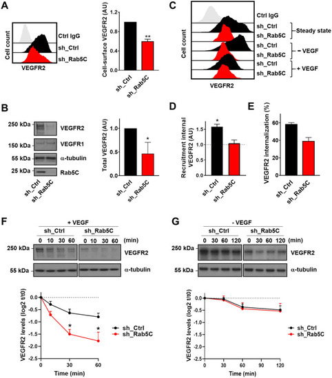
Rab5C protects endosomal VEGFR2 from VEGF-induced lysosomal degradation. a Cell-surface levels of VEGFR2 in sh_Ctrl and sh_Rab5C HUVECs analyzed by flow cytometry. (left) Histograms of a representative experiment, (right) quantification (means + SEM) of 4 independent experiments. Mean fluorescence intensities are expressed relative to those in sh_Ctrl cells. b Western blot analysis (left) of sh_Ctrl and sh_Rab5C HUVEC lysates probed for VEGFR2 and VEGFR1, with α-tubulin serving as a loading control. Blots are representative of 3 independent experiments. Quantification (right) shows the relative VEGFR2 levels normalized to total protein content. Levels in sh_Ctrl cells were set to 1. Results are means + SEM of 3–4 independent experiments. c sh_Ctrl and sh_Rab5C HUVECs were starved for 30 min and then either left untreated or stimulated for 30 min with 50 ng/ml VEGF, whereafter the cell-surface levels of VEGFR2 were analyzed by flow cytometry. d Recruitment of VEGFR2 upon starvation was calculated as the increase in surface levels with respect to those in steady-state. Results are means + SEM of 3 independent experiments. e VEGF-induced VEGFR2 internalization was calculated for sh_Ctrl and sh_Rab5C HUVECs. Mean fluorescence intensities were normalized to steady-state levels. Results are means + SEM of 3–4 independent experiments. f, g HUVECs were starved overnight and subsequently stimulated with 50 ng/ml VEGF (f) or maintained in starvation medium for the indicated times (g). Lysates were subjected to Western blot analysis for VEGFR2, with α-tubulin as a loading control. Blots are representative of 4–5 individual experiments. Quantification of Western blots shows the decline in VEGFR2 levels, expressed relative to the levels at t = 0. Values represent means + SEM of 4–5 independent experiments

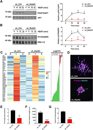
Rab5C regulates endosomal VEGF signaling, the immediate VEGF transcriptome, and sprouting angiogenesis. a Western blot analysis (left) and quantification (right) of phosphorylation of (S473)AKT and total AKT levels at 0, 10, 20, and 30 min of VEGF stimulation (50 ng/ml). b Western blot analysis (left) and quantification (right) of (Y204)ERK-1/2 phosphorylation and total ERK-1/2 levels at 0, 5, 15, and 30 min of VEGF stimulation (50 ng/ml). Representative blots are shown, α-tubulin served as a loading control. Quantifications are means + SEM from 3 independent experiments and expressed relative to t = 0. (c) HUVECs were starved overnight (t = 0), then stimulated with VEGF (50 ng/ml) for 1 h (t = 1) or 4 h (t = 4). VEGF-induced gene expression was then assessed by RNA-seq. Genes have been ordered based on the difference in mean log2 FC between sh_Ctrl and sh_Rab5C. Heatmap shows the corresponding centered and scaled mean expression values for VEGF-induced genes at the indicated time-points. Differences in mean log2 fold change (FC) between sh_Ctrl and sh_Rab5C after 1 h of VEGF stimulation are indicated on the right. Genes that have a higher mean expression in sh_Rab5C with respect to sh_Ctrl are indicated in red, those that have a lower mean expression in green. (d) Sprouting of sh_Ctrl (top) and sh_Rab5C (bottom) HUVECs from collagen-coated beads into fibrin gels. Cells were fixed at 48 h, stained for F-actin (magenta) and nuclei (cyan), and visualized by confocal microscopy. Representative images are shown (maximum projections from z-stacks). Scale bar, 75 μm. e Quantification of the average number of large sprouts/bead. Values represent the means + SEM of 3 independent experiments (20 beads per experiment). f Quantification of the total network length and g the total number of sprouts. A representative experiment is shown. Values represent the means + SEM of n = 16 beads per condition

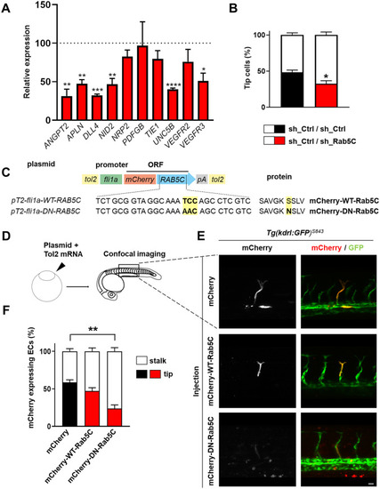
Rab5C is essential for tip cell formation. a sh_Ctrl and sh_Rab5C HUVECs were subjected to qPCR analysis for the indicated tip cell markers. Data are means + SEM of 3–5 individual experiments and are expressed relative to the means in sh_Ctrl cells (indicated by dashed line). b sh_Ctrl and sh_Rab5C cells were differentially labeled using CellTracker dyes, whereafter they were mixed in a 1:1 ratio, adhered to collagen-coated beads, and subjected to VEGF-stimulated sprouting in fibrin gels. After 48 h, cells were fixed and processed for confocal microscopy. Quantification of the number of tip cells was performed by counting the tip cells from confocal z-stacks and normalizing to the total number of cells in that color. Results shown are the means + SEM of 3 pooled independent experiments. Between 8 and 15 beads (containing on average 14 tip cells/bead) were analyzed per condition per experiment. c Schematic of the constructs used for zebrafish experiments. d Constructs encoding mCherry-tagged WT or DN Rab5C were injected together with tol2 transposase mRNA into Tg(kdrl:GFP)s843 zebrafish embryos at the single-cell stage to achieve stable mosaic overexpression in the vasculature. The ISVs were analyzed at 32 hpf. e Maximum projections of z-stacks obtained by confocal microscopy of Tg(kdrl:GFP)s843 embryos expressing mCherry, mCherry-WT-Rab5C, or mCherry-DN-Rab5C, showing the position of positive cells in the ISVs at 32 hpf. Scale bar, 20 μm. f Quantification of the distribution of mCherry, mCherry-WT-Rab5C, or mCherry-DN-Rab5C positive cells. Percentages were calculated per embryo. Shown are the means + SEM. Statistical significance indicates tip cell positioning compared to the mCherry control (mCherry: N = 12 embryos, n = 123 cells; mCherry-WT-Rab5C: N = 17, n = 148; mCherry-DN-Rab5C: N = 14, n = 82)

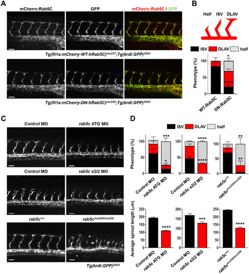
Rab5C is required for sprouting angiogenesis in vivo. a Maximum projections of z-stacks obtained by confocal microscopy showing ISV formation in transgenic Tg(kdrl:GFP)s843 zebrafish embryos stably expressing mCherry-WT-Rab5C (top) or mCherry-DN-Rab5C (bottom). Scale bars, 40 μm. b (top) ISV formation was scored as indicated, (bottom) graph show the results of a representative experiment. Percentages were calculated per embryo (mCherry-WT-Rab5C: N = 10 embryos, n = 72 ISVs; mCherry-DN-Rab5C: N = 12 embryos, n = 88 ISVs). Statistical significance indicates DLAV and ‘half’ phenotypes compared to mCherry-WT-Rab5C. c Maximum projections of z-stacks obtained by confocal microscopy showing ISV formation in Tg(kdrl:GFP)s843 zebrafish embryos injected with Control or rab5c ATG MO (top), Control or rab5c e2i2 MO (middle), or rab5c+/+ versus rab5cmu229/mu229 embryos (bottom). Scale bars, 40 μm. d ISV development (top) as well as average sprout length (bottom) were scored for the indicated conditions at 30–32 hpf (6–10 embryos were analyzed per condition, with 6–8 ISVs per embryo). Statistically significant differences in DLAV and ‘half’ phenotypes, compared to Control MO or rab5c+/+, are indicated

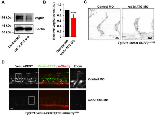
Rab5C controls Vegf and Notch signaling in vivo. a Western blot analysis of zebrafish lysates probed for Vegfr2 with α-actin as a loading control. A representative blot from n = 3 is shown. b Quantification of Western blots showing relative Vegfr2 levels (normalized to α-actin). Levels in embryos injected with Control MO were set to 1. Results are means + SEM (n = 3), 10 embryos were used per condition per experiment. AU, arbitrary units. c Inverted maximum projections of confocal z-stacks acquired by time-lapse microscopy in Tg(fli1a:lifeact-EGFP)mu240 zebrafish embryos injected with Control MO (left) or rab5c MO (right). Scale bar, 10 μm. DA, dorsal aorta. dTg(TP1:Venus-PEST);(kdrl-mCherry)s896 zebrafish embryos demonstrating activation of Notch signaling in embryos injected with Control MOs (top), which is reduced in embryos injected with rab5c MOs (bottom). Scale bar, 20 μm

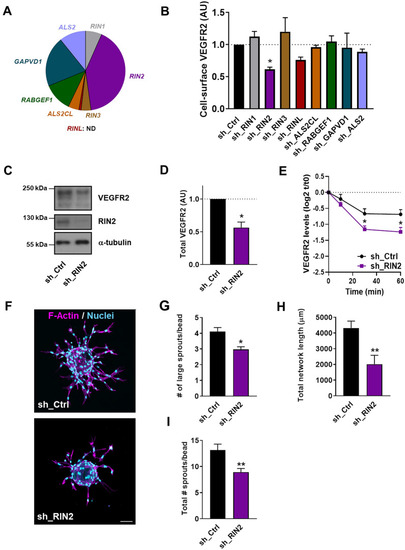
The Rab5 GEF RIN2 protects VEGFR2 from degradation and promotes angiogenic sprouting. a Pie diagram showing the relative mRNA levels of Rab5 GEFs in HUVECs. b Quantification of cell-surface levels of VEGFR2, as assessed by flow cytometry, in HUVECs transduced with shRNAs against the indicated GEFs. Values represent mean fluorescence intensities + SEM of 3–5 independent experiments, expressed relative to sh_Ctrl cells. c Western blot analysis of VEGFR2 and RIN2, using α-tubulin as a loading control. d Quantification of VEGFR2 levels from Western blots. Values represent the means + SEM of 3 individual experiments relative to sh_Ctrl. e Quantification of VEGFR2 degradation from Western blots in sh_Ctrl and sh_RIN2 cells that were starved overnight and subsequently either maintained in growth factor-free medium or stimulated with 50 ng/ml VEGF. Bars represent means + SEM of 3 independent experiments, expressed relative to sh_Ctrl cells at t = 0. f Representative images (maximum projections from confocal z-stacks) showing sprouting of sh_Ctrl and sh_RIN2 HUVECs. Staining shows F-actin (magenta) and nuclei (cyan). Scale bar, 75 μm. g Quantification of the average number of large sprouts/bead for sh_Ctrl and sh_RIN2 cells. Values represent means + SEM of 3 independent experiments (20 beads per experiment). Quantification of h total network length and i average total number of sprouts in sh_Ctrl (n = 18 beads) and sh_RIN2 cells (n = 16 beads). A representative experiment is shown

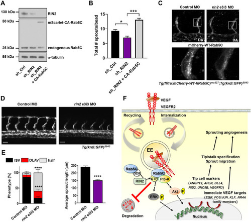
RIN2 regulates Rab5C recruitment and is required for sprouting angiogenesis in vivo. a Representative Western blot showing the expression of RIN2, endogenous Rab5C, and mScarlet-CA-Rab5C in HUVECs. b Quantification of the total number of sprouts/bead was assessed from confocal microscopy z-stacks for the indicated conditions after 24 h of sprouting. Values represent the means + SEM of n = 33 beads (sh_Ctrl), n = 35 beads (sh_RIN2), n = 48 beads (sh_RIN2 + CA-Rab5C), pooled from 3 independent experiments. c Tg(fli1a:mCherry-WT-hRAB5C)mu227;Tg(kdrl:GFP)s843 zebrafish embryos were injected with control MO (left) or rin2 MO (right) and imaged by confocal microscopy to visualize Rab5C localization. Scale bars, 10 μm. DA, dorsal aorta. d Maximum projections of z-stacks obtained by confocal microscopy showing ISV formation at 32 hpf in Tg(kdrl:GFP)s843 zebrafish embryos injected with Control MO (left) or rin2 MO (right). Scale bars, 40 μm. e Quantification of ISV development (left) as well as average sprout length (right) in zebrafish embryos injected with Control MO or rin2 MO. Control MO: N = 12 embryos, n = 84 ISVs; rin2 MO: N = 10 embryos, n = 72 ISVs. Indicated are statistically significant differences in DLAV and ‘half’ phenotypes compared to Control MO. f Model summarizing the main results of this study. Rab5C and the Rab5 GEF RIN2 protect the EE pool of VEGFR2 from VEGF-induced degradation, which sustains VEGF signaling and is required for the expression of immediate VEGF targets and tip cell genes. Together, these events regulate tip cell specification, endothelial cell migration, and sprouting angiogenesis

Acknowledgments
This image is the copyrighted work of the attributed author or publisher, and ZFIN has permission only to display this image to its users. Additional permissions should be obtained from the applicable author or publisher of the image. Full text @ Angiogenesis