The genome structure and protein sequence of zebrafish <italic>cspg4</italic> are similar to other vertebrates.

(A) The genome structure of cspg4 in zebrafish (ENSDART00000112782) compared to human (ENST00000308508), rat (ENSRNOT00000023326.3), mouse (ENSMUST00000035661) and medaka (ENSORLT00000002266.1). Scheme demonstrates the target sites of splicing (MO-e2i2) and translational blocking (MO-start) morpholino (black line) in zebrafish. Gray lines indicate primer pairs used to evaluate pre-mRNA processing. (B) The protein sequence of Cspg4 among zebrafish (UniProt # E7FCN9), rat (UniProt # F1LS79), human (UniProt # Q6UVK1), mouse (UniProt # Q8VHY0) and medaka (UniProt # H2L8T3) were analyzed by Jalview2 software with PAM250 matrix and the Neighbour Joining method [37].

Zebrafish embryo expressed <italic>cspg4</italic> during development.

(A) The expression of cspg4 was detected by RT-PCR from 0 to 120 hpf with actb1 as loading control. (B-H) Expression pattern of cspg4 was revealed by whole-mount in situ hybridization. At 9 (B) and 12 hpf (C), cspg4 was expressed in both the anterior and posterior end of the embryos. Lateral view (D) and dorsal view (E) of 20 hpf embryos showed that cspg4 was mainly expressed in the anterior paraxial and lateral mesoderm (arrow head). (F) Cspg4 was expressed in head, somite, ventral mesenchyme and tail at 24 hpf. Scale bar represents 100 μm. (G) Cspg4 was expressed in head mesenchyme (arrow) and otic placode (asteridsk). (H) Lateral view of 24 hpf embryo showed that cspg4 was expressed in the head mesenchyme (arrow) and pharyngeal region. (I) Cspg4 was expressed in the ventral sclerotome. (J) Cspg4 expressed in jaw, pectoral fins, and trunk at 4 dpf. (K) Ventral view of a 4 dpf larvae showed that cspg4 was expressed in pharyngeal cartilage and the pectoral fin.

<italic>Cspg4</italic> knockdown led to shorter body length.

(A) The 164 bp band indicated the skipping of exon two. (B) Cspg4 knockdown embryos showed shorter body lengths than control morpholino injected embryos at 1 dpf. The body lengths were measured by using the freehand tool in ImageJ (as labeled by the red line). (C) Cspg4 splicing blocking morpholino shortened body length in a dose-dependent manner (each dot represents one embryo; Control-MO, n = 26; 2 ng MO-e2i2, n = 78; 4 ng MO-e2i2, n = 53; 8 ng MO-e2i2, n = 67). (D) Knockdown cspg4 using translational blocking morpholino also shortened body length at 1 dpf (control-MO, n = 140; 1 ng MO-start, n = 60; 2 ng MO-start, n = 139; 4 ng MO-start, n = 105). (E) Combining ineffective doses of cspg4 MO-e2i2 and MO-start shortened the body length at 1 dpf (control-MO, n = 103; 1 ng MO-start +1 ng MO-e2i2, n = 67). In panel (C and D) the significant difference between groups were tested by ANOVA followed by Tukey’s multiple comparison. Different letters (a, b or c) indicate a significant difference between groups (p ≤ 0.05). Student’s t-test was used to analyze data in panel (E). ** indicates p ≤ 0.01. Data are presented as mean ± SD. Scale bar represents 0.5 mm.

PHENOTYPE:
Fish:
Knockdown Reagents:
Observed In:
Stage: Prim-5

<italic>Cspg4</italic> morphants had abnormal pharygeal cartilage at 3 dpf.

The cartilage was stained by using Alcian blue staining. (A, B) The ventral view and lateral view of 4 ng control MO injected embryo at 3 dpf. (C, D) Four ng cspg4 MO-e2i2 injected morphants had disrupted Meckel’s cartilage (arrow head) and underdeveloped ceratohyal cartilage (arrow). (E, F) The phenotypes of abnormal pharyngeal cartilage development were rescued by co-injecting 250 pg cspg4 mRNA with 4 ng MO-e2i2. The number in bottom right indicates the penetrance of phenotype.

<italic>Cspg4</italic> knockdown led to abnormal body axis formation at tailbud stage.

(A) Incomplete body axis organization was caused by cspg4 knockdown. The degrees of angle between anterior end of embryo and tailbud (labeled by yellow line) were measured by ImageJ. (B) Quantitative results of the angle between anterior end and tailbud were analyzed (each dot represents one embryo. Control-MO, n = 97; 2 ng MO-e2i2, n = 78; 4 ng MO-e2i2, n = 52; 8 ng MO-e2i2, n = 66). (C) Knockdown cspg4 using translational blocking morpholino also had larger angle between anterior end of embryo and tailbud (control-MO, n = 144; 1 ng MO-start, n = 70; 2 ng MO-start, n = 122; 4 ng MO-start, n = 73). (D) Co-injecting the ineffective dose of MO-e2i2 (1 ng) and MO-start (1 ng) also led to the same phenotype. (E) The angle between anterior end and tailbud was significantly reduced when co-injecting 250 pg cspg4 mRNA with 4 ng cspg4 morpholino (control, n = 63; knockdown, n = 43; overexpress, n = 99; rescue, n = 51). (F) About 12% (18/148) of embryos had fused eyes (indicated by arrowhead) or only one eye (indicated by arrow). In figure (B), (C) and (E), the significant difference between groups were tested by ANOVA followed by Tukey’s multiple comparison. Different letters (a, b or c) indicate significant difference between groups (p ≤ 0.05). Student’s t-test was used in analyzed data in panel (D), ** indicates p ≤ 0.01. Data are presented as mean ±SD.

Cspg4 transmembrane domain mutant embryo phenocopied <italic>cspg4</italic> overexpression phenotype.

(A) Scheme illustrates wild-type and mutant Cspg4 protein structure. (B) The flag-tag signal of both wild-type and mutant Cspg4 were detected in the cell lysate, but in culture medium the signal of flag-tag was only detected in mutant Cspg4-flag transfected group. (C) Eight percent of cspg4em1twu0405 embryos were cyclopia. (D) Four percent of cspg4em1twu0405 embryos showed midline bifurcation (notochord was indicated by asterisk).

PHENOTYPE:
Fish:
Knockdown Reagents:
Observed In:
Stage: Prim-5

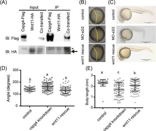
The phenotypes of <italic>cspg4</italic> morphants were rescued by co-injecting <italic>wnt11</italic> mRNA.

(A) Wnt11 had binding affinity with Cspg4. Wnt11-HA (indicated by arrow) was co-precipitated with Cspg4-flag. Asterisk mark (*) indicates non-specific band. (B) The abnormal body axis formation phenotype in cspg4 morphants (4ng MO-e2i2) was rescued by co-injecting 25 pg wnt11 mRNA. Yellow lines indicate the angle between anterior end and tailbud. (C) Co-injecting 25 pg wnt11 mRNA with 4 ng cspg4 morpholino rescued the short body length phenotype. (D) The angle between anterior end and tailbud was reduced in wnt11 rescue group compared to cspg4 knockdown group (control, n = 41; cspg4 knockdown, n = 89; wnt11 rescue, n = 117). (E) The body length of wnt11 rescue group at 1 dpf was significantly longer than cspg4 knockdown group (control, n = 40; cspg4 knockdown, n = 66; wnt11 rescue, n = 109). The significant difference was tested using ANOVA followed by Tukey’s multiple comparison. Different letters (a, b or c) indicate significant difference between groups (p ≤ 0.05). Data are presented as mean ± SD.

PHENOTYPE:
Fish:
Knockdown Reagent:
Observed In:
Stage Range: Bud to Prim-5

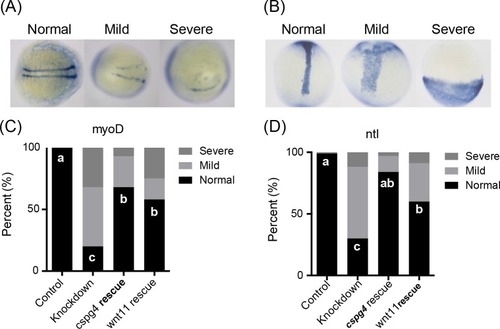
Knockdown of <italic>cspg4</italic> affects paraxial mesoderm marker <italic>myoD</italic> and notochord marker <italic>ntl</italic> expression pattern.

(A) Cspg4 morphants had a wider and shorter (mild) or unilateral (severe) myoD expression pattern compared to control morphants (normal) at 12 hpf. (B) The notochord was wider and shorter (mild) or absence (severe) in cspg4 morphants compared to control morphants (normal) at 12 hpf. (C) The percentage of embryos with abnormal myoD expression pattern in each group (control, n = 41; cspg4 knockdown, n = 40; cspg4 rescue, n = 28; wnt11 rescue, n = 24). (D) The percentage of embryos with abnormal ntl expression pattern in each group (control, n = 73; cspg4 knockdown, n = 43; cspg4 rescue, n = 67; wnt11 rescue, n = 35). The significant difference of normal rate was tested using Kruskal–Wallis test followed by Dunn’s multiple comparisons test. Different letters (a, b or c) indicate significant difference of normal rate between groups (p ≤ 0.05).

EXPRESSION / LABELING:
Genes:
Fish:
Knockdown Reagent:
Anatomical Terms:
Stage: 5-9 somites
PHENOTYPE:
Fish:
Knockdown Reagent:
Observed In:
Stage: 5-9 somites
Acknowledgments
This image is the copyrighted work of the attributed author or publisher, and ZFIN has permission only to display this image to its users. Additional permissions should be obtained from the applicable author or publisher of the image. Full text @ PLoS One