FIGURE SUMMARY
Title

Spatio-temporal expression and distribution of collagen VI during zebrafish development

Authors
Tonelotto, V., Trapani, V., Bretaud, S., Heumüller, S.E., Wagener, R., Ruggiero, F., Bonaldo, P.
Source
Full text @ Sci. Rep.

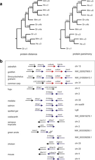
Phylogenetic analysis of zebrafish ColVI genes. (a) Phylogenetic trees of the ColVI α4, α5 and α6 chains in different species, obtained by amino acid sequence comparison of the regions spanning the C1 and C2 domains in the corresponding α chains. The sequences from zebrafish (z), mouse (m) and human (h) were aligned using the PILEUP program of the GCG package, using default parameters. The trees were constructed using the PROTEIN PARSIMONY, PROTEIN DISTANCE, FITCH and CONSENSE tools of the PHYLIP package version 3.69. Bootstrap analyses using 100 replicates were performed to show the significance. Numbers indicate the statistical weight of the individual branches. The C1 and C2 domains of zebrafish ColVI α1 chain were used as outgroup. (b) Comparative maps of syntenic regions of the zebrafish col6a4b gene. Genes encoding the corresponding α4 chain in different species are indicated by red arrows. Neighboring syntenic genes are indicated by blue arrows. For simplicity, the orientation of the genes on the chromosomes was adjusted to that of zebrafish chromosome 13. Members of the carp family (Cyprinidae) are boxed.

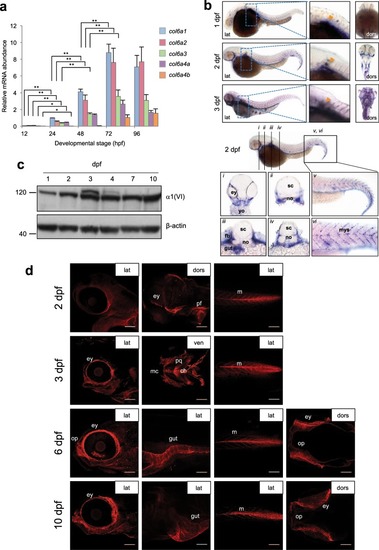
ColVI expression in zebrafish at different developmental stages. (a) qRT-PCR for col6a1, col6a2, col6a3, col6a4a and col6a4b transcripts in zebrafish embryos and larvae from 12 hpf to 96 hpf. Data were normalized to arp. The expression levels of the different genes were compared to col6a1 expression at 24 hpf, which was arbitrarily set to 1, and represent the mean of at least three independent experiments. Error bars indicate s.e.m. (**P < 0.01). (b) Whole-mount in situ hybridization for col6a1 in zebrafish embryos and larvae. The top panels show lateral (lat) and dorsal (dors) views of 1- to 3-dpf embryos labeled with the col6a1 probe. The bottom panels show transverse sections at different head and trunk levels (i-iv, as indicated on the lateral view at the top) and higher magnifications of the tail region (v and vi) of 2-dpf embryos labeled with the col6a1 probe. At 1 dpf, labeling for col6a1 mRNA is restricted to two lines of slow muscle fibers and to the pectoral fin buds (orange arrowhead in the magnified area highlighted by the dotted box). At 2 and 3 dpf, strong signals for col6a1 transcript are detected in vertical myosepta, pectoral fins (orange arrowhead in the magnified areas highlighted by the dotted boxes), branchial arches, axial vasculature and gut. All panels were cropped from the background. (c) Western blot for α1(VI) chain in protein extracts from zebrafish embryos and larvae at the indicated stages. β-actin was used as a loading control. Numbers on the left indicate sizes (in kDa) of protein standard markers. The extra-band observed at 3 and 4 pdf may correspond to post-translational modifications. The cropped blots for α1(VI) chain and β-actin derive from different parts of the same gel. (d) Whole-mount immunofluorescence labeling with anti-ColVI antibody in zebrafish larvae at different developmental stages from 2 dpf to 10 dpf, as indicated. The panels show lateral (lat), ventral (ven) and dorsal (dors) views of different head and trunk regions. From 2 dpf onwards, ColVI immunoreactivity (red) is present in the connective tissue surrounding the eye, in the pectoral fins and in myosepta. At 3 dpf, ColVI deposition extends to craniofacial elements and intestine. From 6 dpf onwards, ColVI labeling is also detected in the connective tissue surrounding olfactory pits. Scale bar, 100 µm. ch, ceratohyal; ey, eye; fb, fin buds; m, myosepta; mc, Meckel’s cartilage; myoseptum, mys; no, notochord; op, olfactory pit; pf, pectoral fins; pq, palatoquadrate; sc, spinal cord; yo, yolk.

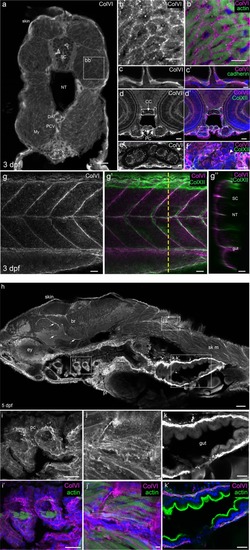
Characterization of ColVI deposition in 3- and 5-dpf developing zebrafish. (a-g’) Immunofluorescence images of transverse sections (af) or whole-mount (g-g”) 3-dpf larvae at the level of the trunk. Samples were labeled with anti-ColVI antibodies (panels a-e, g in grey; panels b’,c’,d’,f,g’,g” in magenta) and, where indicated, with phalloidin to reveal actin (green; panel b’), or with antibodies against the epithelial marker cadherin (green; panel c’) and the mesenchymal marker collagen XII (green; panels d’,f,g’,g”). Nuclei were stained with Hoechst (blue; panels b’,c’,d’,f). (a) Global view of ColVI immunoreactivity at the level of the trunk. (b,b’) Zoomed images of the boxed region in (a), showing myotomal muscle cells. (c,c’) Epidermis. (d,d’) Transverse section of the head. (e,f) Zoomed images of the boxed regions of panels d and d’, showing chondrocyte stacks in the cranial cartilage. White arrowhead points to intracellular ColVI staining in chondrocytes. (g) Image of vertical myosepta, obtained from a z-projection of confocal stack of lateral views. (g’) Merge image of the same acquisition, showing ColVI and collagen XII double staining. (g”) Orthogonal view at the level indicated by the yellow dashed line in g’. (h-k’) Immunofluorescence images of sagittal sections of 5-dpf larvae. Samples were labeled with α1(VI) antibodies (grey or magenta) and, where indicated, with phalloidin (green; panels i’,j’,k’). Nuclei were stained with Hoechst (blue; panels i’,j’,k’). (h) Global view of ColVI immunoreactivity. Arrows indicate ColVI-positive vessels. (i-k’) Zoomed images of the boxed regions of panel h. (i,i’) Ceratobranchial cartilage. (j,j’) Skeletal muscle at the trunk level. (k,k’) Intestine. Scale bars, 50 μm (h); 25 μm (d,d’,g-g”,i,i’,k,k’); 20 μm (a); 10 μm (b,b’,e,f); 5 μm (c,c’,j, j’). br, brain; CC, chondrocranium; DA, dorsal aorta; ey, eye; li, liver; My, myotome; NT, notochord; pc, pharyngeal cartilage; PCV, posterior cardinal vein; PC, pharyngeal cartilage; pf, pectoral fin; SC, spinal cord; sk m, skeletal muscle.

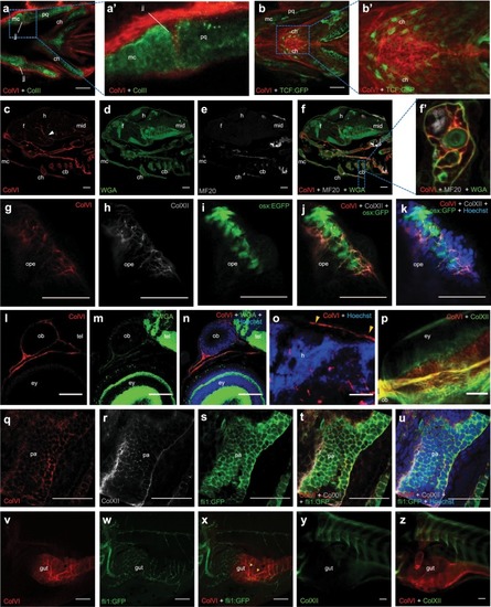
Spatio-temporal pattern of ColVI distribution in larvae from wild-type animals and transgenic reporter fish lines. (a-a’) Individual z-stack of whole-mount immunofluorescence for ColVI (red) and collagen II (green) in 3-dpf larvae. Ventral view. A strong ColVI labeling in evident in the connective tissue surrounding craniofacial cartilages and in the jaw joint. Panel a’ is a magnification of the dotted area of panel a, showing the deposition of ColVI in the jaw joint. (b-b’) Confocal z-stacks of whole-mount immunofluorescence for ColVI (red) in 6-dpf Tg(7xTCF-Xla.Siam:GFP)ia4 (TCF-GFP, green) larvae, showing ColVI labeling near Wnt-positive cells in craniofacial cartilages. Ventral view. Panel b’ is a magnification of the dotted area of panel b, showing ColVI labeling in close apposition to Wnt-positive cells in the ceratohyal cartilages. (c-f’) Section of 7-dpf larvae stained with anti-α1(VI) antibodies (red), WGA (green) and anti-MF20 antibodies (gray). ColVI labeling is present in craniofacial cartilaginous elements, as well as in blood vessels (arrowhead). Panel f’ is a magnification of the dotted area of panel f, showing ColVI labeling in the perichondrium surrounding the cartilaginous elements of the pharyngeal arches, as revealed by co-localization with WGA. (gk) Individual z-stack of whole mount immunostaining of 6-dpf Tg(osx:nuGFP) (osx:GFP, green) larvae operculum, stained with ColVI (red) and collagen XII (gray) antibodies and Hoechst (blue). ColVI and collagen XII surround operculum osteoblasts. (ln) Section of a 7-dpf larva stained with anti-ColVI antibodies (red), WGA (green) and Hoechst (blue). ColVI labeling is present in the connective tissue around brain, olfactory pit and eye. (o) Section of a 7-dpf larva stained with anti-ColVI antibodies (red) and Hoechst (blue), showing ColVI labeling in the meninges (arrowhead). (p) Confocal z-stacks of whole-mount immunofluorescence for ColVI (red) and collagen XII (green) on 6 dpf larvae eye, showing ColVI deposition in the connective tissue surrounding the eye. Dorsal view. (qu) Individual z-stack of whole mount immunostaining for ColVI (red), collagen XII (gray) antibodies and Hoechst (blue) in 2-dpf Tg(fli1:EGFP) (fli:EGFP, green) larvae at the level of pharyngeal arches. ColVI and collagen XII are part of the bordering tissue of migrating pharyngeal pouches. (vx) Confocal z-stacks of whole-mount immunofluorescence for ColVI (red) in 6-dpf Tg(fli1:EGFP) (fli:EGFP, green) larvae. A strong pattern of ColVI labeling is found around blood vessels (arrowheads) of the intestine. (y-z) Confocal z-stacks of whole-mount immunofluorescence for ColVI (red) and collagen XII (green) in 6-dpf larvae, showing that ColVI but not collagen XII is deposited in the intestine. Scale bar, 100 µm (vx) or 50 µm (au,y,z). cb, ceratobranchial; ch, ceratohyal; ey, eye; f, forebrain; h, hindbrain; jj, jaw joint; mc, Meckel’s cartilage; mid, midbrain; op, olfactory pit; ope, operculum; pa, pharyngeal arch; pq, palatoquadrate.

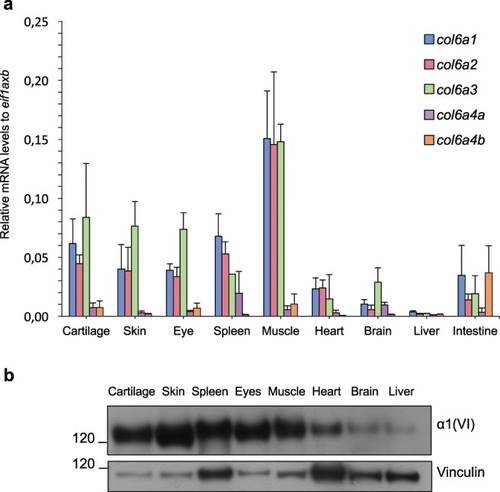
ColVI expression in adult zebrafish tissues. (a) qRT-PCR for col6a1, col6a2, col6a3, col6a4a and col6a4b transcripts in different tissues of adult (8- to 12-mpf) fish. Data were normalized to eif1axb expression. Error bars indicate s.e.m. (b) Western blotting for α1(VI) chain in protein extracts of adult (8- to 12-mpf) fish tissues. Vinculin was used as a loading control. Number on the left indicate sizes (in kDa) of protein standard markers. The cropped blots for α1(VI) chain and vinculin derive from different parts of the same gel.

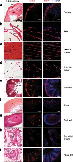
Characterization of ColVI deposition in adult zebrafish. (ai) Sagittal sections of adult (8-to 12-mpf) fish tissues analyzed by haematoxylin-eosin staining (H&E, left panels) and by immunofluorescence labeling with anti-ColVI antibodies (red, middle and right panels). Nuclei were stained with Hoechst (blue, right panels). Scale bar, 50 µm. a, adipocyte; bs, bony scale; cb, ceratobranchial; ce, ceratohyal; cr, cryptae; endom, endomysium; en, endothelium; ep, epithelium; gc, goblet cell; ir, iris; me, mucosal epithelium, pl, primary lamella; sl, secondary lamella; st, stroma; te, telencephalon.

ZFIN is incorporating published figure images and captions as part of an ongoing project. Figures from some publications have not yet been curated, or are not available for display because of copyright restrictions.

Acknowledgments
This image is the copyrighted work of the attributed author or publisher, and ZFIN has permission only to display this image to its users. Additional permissions should be obtained from the applicable author or publisher of the image. Full text @ Sci. Rep.