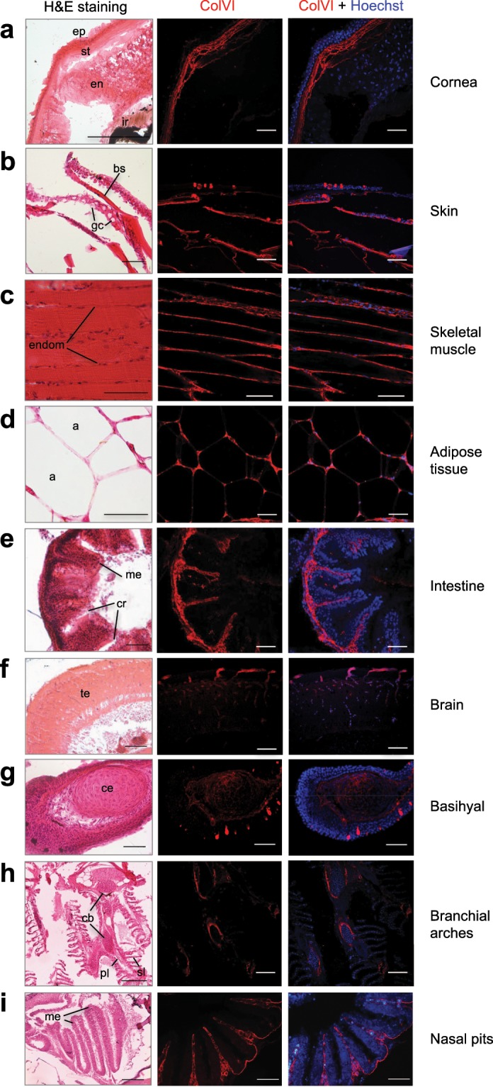
IMAGE

Figure 6

ID
ZDB-IMAGE-200102-8
Antibodies
Source
Figures for Tonelotto et al., 2019
Image
Figure Caption

Figure 6

Characterization of ColVI deposition in adult zebrafish. (ai) Sagittal sections of adult (8-to 12-mpf) fish tissues analyzed by haematoxylin-eosin staining (H&E, left panels) and by immunofluorescence labeling with anti-ColVI antibodies (red, middle and right panels). Nuclei were stained with Hoechst (blue, right panels). Scale bar, 50 µm. a, adipocyte; bs, bony scale; cb, ceratobranchial; ce, ceratohyal; cr, cryptae; endom, endomysium; en, endothelium; ep, epithelium; gc, goblet cell; ir, iris; me, mucosal epithelium, pl, primary lamella; sl, secondary lamella; st, stroma; te, telencephalon.

Figure Data
Acknowledgments
This image is the copyrighted work of the attributed author or publisher, and ZFIN has permission only to display this image to its users. Additional permissions should be obtained from the applicable author or publisher of the image. Full text @ Sci. Rep.