- Title
-
Collagen triple helix repeat containing 1a (Cthrc1a) regulates cell adhesion and migration during gastrulation in zebrafish
- Authors
- Cheng, X.N., Shao, M., Shi, D.L.
- Source
- Full text @ Exp. Cell Res.
|
Knockdown of cthrc1a disrupts epiboly and axis extension. (A–J) Representative live images of CoMO-injected and cthrc1MO-injected embryos. The white triangle marks the embryonic shield; red arrows indicate the blastoderm margin, and red arrowheads show the anterior tip of hypoblast; yellow lines define the angle formed between the dorsal side and the anterior tip of hypoblast, with vertex at the geometric centre of the embryo, which represents the extent of animal pole migration of the anterior tip of hypoblast. Embryos at 5.5 hpf, 7.5 hpf, and 10 hpf are positioned with animal pole on the top and dorsal region on the right, and embryos at 14 hpf and 26 hpf are lateral views. (K–P) In situ hybridisation of CoMO-injected and cthrc1MO-injected embryos using indicated markers, showing defective CE movements. Dorsal view for all embryos oriented anterior region up. (Q–V) Rescue of axis extension defects in cthrc1a morphants by myc-Cthrc1a at 10 hpf and 27 hpf. (W) Statistical analysis of the extent of hypoblast migration in embryos injected with CoMO, cthrc1MO, or coinjected with myc-cthrc1a and cthrc1MO. Bars represent the mean ± s.d. from three independent experiments (***, P < 0.001). Scale bars: (A-D, F–I, K–P, Q, S and U), 200 μm; (E, J, R, T and V), 500 μm. PHENOTYPE:
|

ZFIN is incorporating published figure images and captions as part of an ongoing project. Figures from some publications have not yet been curated, or are not available for display because of copyright restrictions. PHENOTYPE:
|
|
Knockdown of cthrc1a impairs CE movements. (A–D) Convergence of lateral cells in CoMO-injected and cthrc1MO-injected embryos. A small group of lateral margin cells were labelled at 6 hpf, and their dorsal-ward convergence was examined at 10 hpf. (E) Plot compares the extent of convergence of labelled cells in CoMO-injected and cthrc1MO-injected embryos. Bars represent the mean ± s.d. from three independent experiments (***, P < 0.001). The inset image is an animal pole view showing the angle formed between labelled cells and the line perpendicular to the dorsoventral axis, with green dot representing the centre of the image. (F–I) Anteroposterior extension of midline cells in CoMO-injected and cthrc1MO-injected embryos, by following the movement of a small group of cells initially labelled at shield region. (J) Plot compares the extent of extension of labelled cells in CoMO-injected and cthrc1MO-injected embryos. The inset image shows the angle formed between the dorsal side and most anteriorly labelled cells. Bars represent the mean ± s.d. from three independent experiments (***, P < 0.001). (K) Injection of cthrc1MO along with RLDx in the blastomeres on the right side. (L) Anteroposterior extension defect on the injected side, as shown by the curved axis (dashed line). Scale bars: (A-D, F–I and K), 200 μm; (L) 250 μm. PHENOTYPE:
|

ZFIN is incorporating published figure images and captions as part of an ongoing project. Figures from some publications have not yet been curated, or are not available for display because of copyright restrictions. PHENOTYPE:
|
|
Functional interaction between Cthrc1a and integrin β1 in axis extension. (A) Embryos were injected with CoMO, low dose of dnß1 mRNA, low dose of cthrc1MO, or a mixture of cthrc1MO and dnß1 mRNA at 1-cell stage, statistical analysis of the extent of hypoblast migration in different injected embryos was performed at bud stage, as shown in the inset image. A total of 12 embryos randomly selected from 3 independent experiments were analysed for each condition. Numbers in each column represent total embryos injected. The results are expressed as mean ± s.d. from three independent experiments (*, P < 0.05; **, P < 0.01; Student's t-test). (B–E) Representative phenotypes for each condition at 27 hpf, as indicated. Scale bar: (B–E) 500 μm. |

ZFIN is incorporating published figure images and captions as part of an ongoing project. Figures from some publications have not yet been curated, or are not available for display because of copyright restrictions. PHENOTYPE:
|
|
Temporal and spatial expression patterns of cthrc1a during early development. (A) RT-PCR analysis of cthrc1a temporal expression at indicated stages. ß-actin was used as loading control. (B) At 6 hpf, cthrc1a transcripts are localised at the entire margin. Animal pole view. (C) At 8 hpf, dorsal view shows cthrc1a expression in the dorsal midline and lateral regions. (D) Lateral view of a gastrula at 10 hpf, cthrc1a expression is localised in the dorsal and lateral regions along the anteroposterior axis. (E) Cross section of a shield stage embryo through marginal zone. Dorsal midline is placed upwards, and arrow shows cthrc1a expression in the embryonic shield. (F) Sagittal section of an embryo at 80% epiboly showing enhanced staining in the dorsal yolk syncytial layer and the dorsal midline hypoblast cells. Animal pole region up and dorsal region on the right. Scale bars: 200 μm. |
|
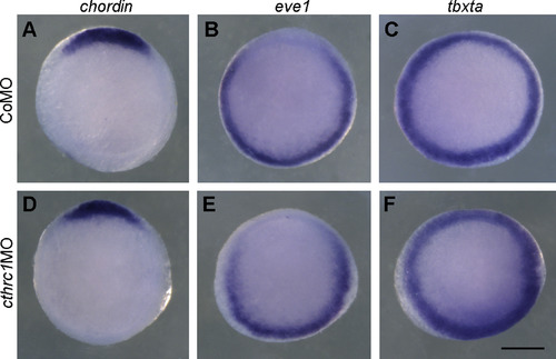
Expression of dorsoventral markers at shield stage following cthrc1aknockdown. Dorsal (chordin), ventral (eve1), and pan-mesoderm (tbxta) markers are indicated on the top. (A-C) CoMO-injected embryos. (D-F) Translation-inhibiting cthrc1MO-injected embryos. Animal pole view, with dorsal region on the top. Scale bar: 200 μm.2 |

ZFIN is incorporating published figure images and captions as part of an ongoing project. Figures from some publications have not yet been curated, or are not available for display because of copyright restrictions. PHENOTYPE:
|

ZFIN is incorporating published figure images and captions as part of an ongoing project. Figures from some publications have not yet been curated, or are not available for display because of copyright restrictions. PHENOTYPE:
|
|
Overexpression of Squint converts all blastoderm cells into mesendoderm. Analysis by in situ hybridisation of ntl expression in indicated embryos at shield stage. (A) A CoMO-injected embryo. (B) A CoMO-injected embryo overexpressing Squint. (C) A cthrc1MO-injected embryo overexpressing Squint. Scale bar: 200 μm.6 |
|
Cell non-autonomous function of Cthrc1a on cell migration. A small group of RLDx-labelled cells from a CoMO-injected embryo or a cthrc1MO-injected embryo were transplanted to the dorsal margin of a normal embryo at 6 hpf, and the distribution of labelled cells was followed at 10 hpf. (A and B) Normal embryos transplanted with CoMO-injected cells. (C and D) Normal embryos transplanted with cthrc1MO-injected cells. Both CoMO-injected and cthrc1MO-injected cells migrate to similar position in normal host embryos at the end of gastrulation. Scale bar: 200 μm.8 |


