- Title
-
Pathological mineralization in a zebrafish enpp1 mutant exhibits features of Generalized Arterial Calcification of Infancy (GACI) and Pseudoxanthoma Elasticum (PXE)
- Authors
- Apschner, A., Huitema, L.F., Ponsioen, B., Peterson-Maduro, J., Schulte-Merker, S.
- Source
- Full text @ Dis. Model. Mech.
|
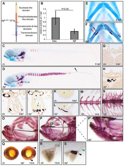
dgfhu4581 mutants show decreased phosphodiesterase activity and multiple ectopic calcifications. (A) Depiction of the Enpp1 protein structure; the dgfhu4581 allele represents a frame shift at amino acid 331, leading to a premature stop codon. (B) Phosphodiesterase I activity is significantly reduced in the lysate of dgf embryos. Means±1 s.d. are shown. Sib(s), wild-type sibling(s). Alizarin-Red (staining mineralized tissue) and Alcian-Blue (staining cartilage) staining of a sibling embryo (C) and dgf mutant (D) at 8 dpf showing extensive ectopic calcification of the notochord, as well as calcification of the neural tube (D, arrowhead). (E) Ventral view of ceratohyal cartilage element of sibling embryo; in mutant embryos, early onset of perichondral ossification (F, red arrowhead), as well as spots of ectopic cartilage calcification (F, black arrowhead), were observed. van Kossa (brown, staining mineralized tissue) and van Gieson (red, staining osteoid) staining on transverse sections of the brain of a sibling (G) and a dgf mutant with intracranial calcification (H). Transverse section through the heart region of sibling (I) and mutant (J) embryos, both displaying mineralized cleithra (cl) and basobranchial (bb). Mutants (J) in addition display ectopic mineralization between myocard and epicard (red arrowhead) and within the heart (blue arrowhead). (K) Transverse section at the level of the yolk sac of a sibling; (L) the mutant displays ectopic mineralization of the skin. Axial skeleton at the level of the dorsal fin of a sibling (M) and mutant (N) 4-week-old (4wk) fish. Mutants display not only fusion of vertebral bodies but also of neural and haemal arches (N). Alizarin-Red staining of juvenile sibling (O) and mutant (P and enlarged image of the indicated area in P2). Note the ectopic mineralization at the ethmoid plate cartilage element (green arrowhead in P) and nodules of mineralization at the dentary (black arrowheads in P,P2). (Q) Alizarin-Red staining showing ectopic mineralization (black arrowheads) surrounding the eye of a dgf adult mutant (also green arrowhead in P). (R) In the heart of adult zebrafish, no mineralization was visible in siblings. (S) In mutants extensive ectopic calcification was found upon Alizarin-Red staining in the bulbus arteriosus (black arrowhead) but not in the ventral aorta (green arrowhead). Bb, basobranchial; Cl, cleithrum; y, yolk. PHENOTYPE:
|
|
Soft tissue calcifications in dgf mutants probably represent passive calcium depositions. collagen10a1:YFP (col10) transgene in sibling (A) and mutant (B). Note that no expression of collagen10a1 was detected at sites of ectopic mineralization (Alizarin Red) at the heart (B, blue arrow). osteocalcin:GFP (osc) combined with Alizarin staining in siblings (C) and dgf mutants (D), no ectopic expression of osc was observed to colocalize with ectopic mineralization in the heart region and pectoral fin (D, blue arrows). Calcein staining marks calcifications in collagen2a1a:mCherry (col2) transgenic line in wild-type siblings (E) and dgf mutants (F). The dgf mutant shows ectopic calcifications in the cranium (F, white arrows), however no ectopic expression of collagen2a1a was observed. Alizarin staining and collagen10a1:YFP transgene expression in the axial skeleton of a sibling (G) and dgf embryo (H), ectopic mineralization of the notochord sheet occurs independently of collagen10a1 expression (white arrows, H). In situ hybridization for spp1 (blue) in siblings (I,K) and dgf mutants (J,L). Note upregulation of spp1 in mutant bone elements. Further ectopic expression occurs at loci that are frequently affected by ectopic mineralization in mutants (arrowheads in J,L; compare with supplementary material Fig. S1H). cl, cleithrum; ps, parasphenoid; sib, sibling. |
|
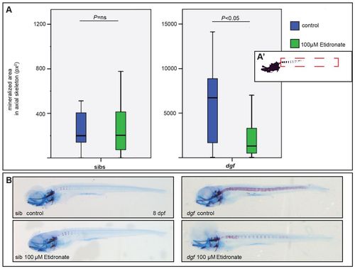
Treatment with the pyrophosphate analog etidronate rescues aspects of the dgf phenotype. (A) Measurements (in pixel area) of the mineralized area in the axial skeleton of Alizarin-Red- and Alcian-Blue-stained siblings (sibs; left panel) and mutants (right panel), which were either untreated (blue) or treated with 100 μM Etidronate (green). A2 indicates the region of interest that was measured for the analysis. No significant difference (ns) occurs in siblings (A,B left panels); in treated dgf mutants, the mineralized area was significantly reduced when compared with untreated dgf mutants (A,B right panels). n=50 (dgf 100 μM Etidronate); n=48 (dgf control); n= 24 (siblings 100 μM Etidronate); n=24 (siblings control). PHENOTYPE:
|
|
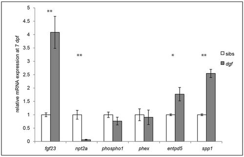
Expression of regulators of phosphate and biomineralization is perturbed in dgf mutants. qPCR analysis showing the relative gene expression levels of genes involved in phosphate homeostasis and biomineralization in siblings (sib) compared with those in dgf mutants, normalized to the expression of ef2a. *P≤0.05, **P≤0.01. Means ± s.e.m. are shown. |
|
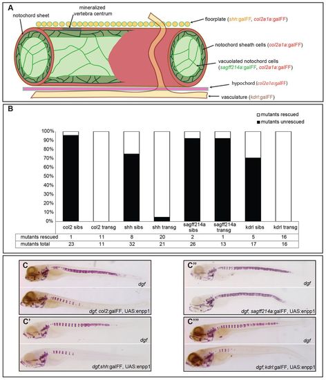
Distal expression of enpp1 is sufficient to prevent ectopic calcifications in the notochord. (A) Overview of the tissue-specific lines that were used in the different rescue experiments. (B) Analysis and quantification of dgf embryos and dgf transgenic lines with respect to the notochord phenotype (shown as percentages of the total number of embryos examined). Sibs, siblings; transg, transgenic line. (C–C4) Representative examples of non-transgenic dgf embryos and their transgenic dgf siblings for the respective constructs after Alizarin-Red staining. |
|
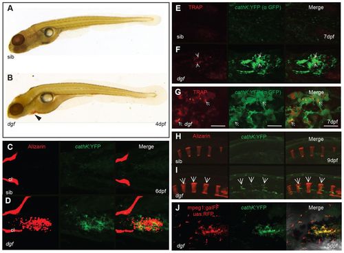
Cells expressing osteoclastic markers appear at ectopic mineralization sites in dgf mutants. Staining of Trap in a wild-type sibling (A) and dgf embryo (B). Trap staining (red) was visible in a dgf embryo at the region of the heart and yolk sac (B, arrowhead). Ventral view of sibling (C) and mutant (D) embryo with ectopic mineralization in the heart and yolk sac region. (C) No cathepsinK-positive cells were visible in siblings at this timepoint; (D) mutants showed colocalization of ectopic soft tissue mineralization and cathepsinK-positive cells, but no cathepsinK-positive cells were aligned to skeletal elements, such as the cleithrum (cl). (E,F) Ventral view of the yolk sac area of embryos that had been stained for Trap and with an antibody against GFP (α GFP). (F) Trap staining appeared in association with cathepsinK-positive cells. White arrows indicate loci of osteoclasts with high Trap activity. (G) Higher-magnification image of cathepsinK and Trap colocalization in a dgf mutant. Scale bar: 20μm. The staining of Trap in E–G is pseudo-colored for improved visibility. In the axial skeleton, cathepsinK-positive cells were not visible in siblings (H); however, in dgf mutants (I), cathepsinK-positive cells appeared from 9 dpf onwards and colocalized with mineralized vertebral bodies. White arrows indicate osteoclasts aligning with vertebral elements. (J) Ventral view of accumulating cathepsinK-positive cells in the skin of the heart region. These cells also express the macrophage marker mpeg1. |
|
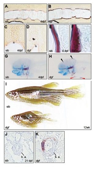
Van kossa/van Gieson staining shows segmented mineralization of the notochord sheath in siblings (A), in dgf mutants show ectopic calcification of the intervertebral spaces (B). Transverse section through the neuraltube of sibling (C) and mutant with ectopic calcification (D). Alizarin red/Alcian blue stained cleithrum and pectoral fin cartilage of siblings (E) and dgf mutant (D) showing ectopic calcification. Overview of Alizarin red/Alcian blue stained sibling (G) and mutant (H). The black arrowheads indicate cranial calcifications, the red arrowheads point at mineralizations of the skin surrounding the yolk sac and heart (H). dgf mutants can reach adulthood in rare cases but remain smaller (I). Transverse section of alizarin stained juvenile embryos at the bulbus arteriosos (b. a.), no mineralization is visible in silbing (J), circumferential calcification in the dgf mutant (K). PHENOTYPE:
|
|
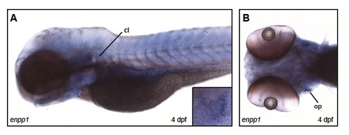
Lateral (A) and ventral (B) view of in-situ hybridisation showing the expression pattern of enpp1 at 4 dpf. Note: expression in bone elements such as cleithrum (cl) (A) and the opercle (op) (B, box in A). |

