- Title
-
odd-skipped related 2 is required for fin chondrogenesis in zebrafish
- Authors
- Lam, P.Y., Kamei, C.N., Mangos, S., Mudumana, S., Liu, Y., and Drummond, I.A.
- Source
- Full text @ Dev. Dyn.
|
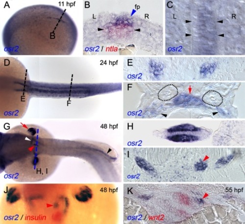
osr2 expression during zebrafish development. A: At 11 hpf, osr2 is expressed in the posterior embryonic midline (tilted lateral view, anterior to the left). B: Cross-section of an 11-hpf embryo at the level of the dotted line indicated in A showing osr2 (blue arrowhead) expression in floorplate (fp) cells overlying the notochord marked by red ntl expression (black arrowheads). C: Expression of osr2 is biased to cells on the right side of the forming floorplate. Black arrowheads mark the boundaries of the notochord. L, left; R, right. D: At 24 hpf, osr2 is expressed in the pronephric proximal tubule and gut. E, F: Cross-section of 24 hpf embryo indicated in Figure 1D showing osr2 expression in the proximal pronephros (E) and gut (F; red arrow). Distal tubules of the pronephros do not express osr2 (dotted circles). osr2 expression is also observed in mesenchyme cells (black arrowheads) ventral and lateral to the pronephros. G: At 48 hpf, osr2 is expressed in fin (red arrow), pronephric proximal tubule (white arrowhead), posterior gut (black arrowhead), and in gut mesenchyme (red arrowhead) at the level for the pectoral fin. H, I: Cross-section of the embryo in G at the level indicated showing expression of osr2 in pectoral fin mesenchyme (H) pronephric tubules, and gut mesenchyme (I; red arrowhead). J: Double in situ hybridization showing gut mesenchyme (red arrowhead) expressing osr2 (blue) anterior to the exocrine pancreas as indicated by insulin (red) expression at 48 hpf. K: Gut mesenchyme (red arrowhead) expressing osr2 (blue) was adjacent to the pneumatic duct as indicated by wnt2 (red) expression at 55 hpf. EXPRESSION / LABELING:
|
|
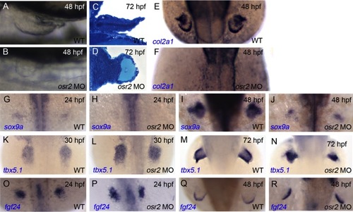
osr2 knock down results in abnormal pectoral fin. Brightfield images of control fins at 48 hpf show the pectoral fin extending toward the posterior (A) while osr2 morphants display only a fin stump (B). Embryos are oriented with anterior to the left and dorsal to the bottom in A, B. C: Methylene blue–stained transverse section of normal pectoral fin at 72 hpf. D: osr2 morphants showed abnormal pectoral fin morphology and lacked differentiated chondrocytes. col2a1 in situ hybridization analysis at 48 hpf of control embryos (E) and osr2 morphants (F). osr2 morphants showed reduction or disappearance of col2a1 expression in the pectoral fin. G–J: sox9a expression was reduced in osr2 morphants at both 24 hpf (H) and 48 hpf (J) compared to controls (G, I). K–N: tbx5a expression in lateral mesoderm at 30 hpf (K, L) and in the pectoral fin buds at 72 hpf (M, N) was not affected in osr2 morphants compared to controls. O–R: fgf24 expression is unaffected by osr2 knock down and is unchanged in the lateral mesoderm at 24 hpf (O, P) and is restricted to the apical ectodermal ridge in both control and osr2 morphants at 48 hpf (Q, R). Embryos in E, F, G–R are displayed from a dorsal view and anterior to the top. EXPRESSION / LABELING:
PHENOTYPE:
|
|
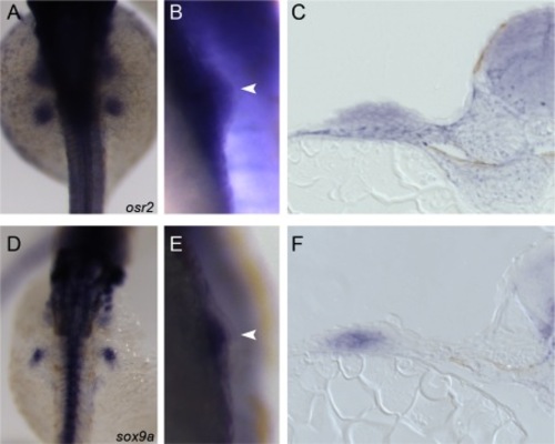
osr2 and sox9a are coexpressed in the developing fin bud. Whole-mount in situ hybridization at 32 hpf shows that both osr2 (A) and sox9a (D) are expressed in the lateral plate in the region of the newly forming fin bud. osr2 (B) appears to be expressed throughout the fin bud, while sox9a (E) shows a more restricted expression. Transverse sections through the center of the fin bud show that osr2 is expressed uniformly throughout the fin bud mesenchyme (C), while sox9a-expressing chondrocytes are restricted to the center (F). Embryos in A, D are displayed from a dorsal view with anterior to the top. In B, E, arrowhead indicates fin bud with proximal to the left. EXPRESSION / LABELING:
|
|
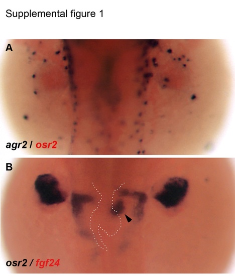
osr2 expression in gut-adjacent mesenchyme. A: Double whole-mount in situ hybridization of a 48-hpf embryo showing gut morphology (agr2; blue-black) and osr2 expression (light red). B: Double in situ hybridization with osr2 probe (blue-black) and fgf24 (light red) showing osr2 expression adjacent to the gut at the approximate position of the future pneumatic duct (arrowhead). Gut morphology from A superimposed in white dashed line in B. |
|
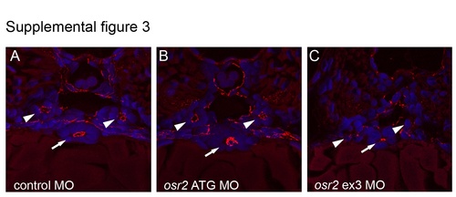
Epithelial development and polarity is not affected in osr2 morphants. A: Histological section of a control embryo at the level of the mid-trunk shows cross-sections of the kidney (arrowheads) and gut (arrow) stained with anti-ZO-1, highlighting polarized apical cell–cell junctions. Both osr2 ATG morphants (B) and osr2 exon3 splice morphants (C) also show normal epithelial polarized junctions in the kidney and gut. Blue, DAPI stained nuclei. Representative sections from n=3 for each morpholino. |
|
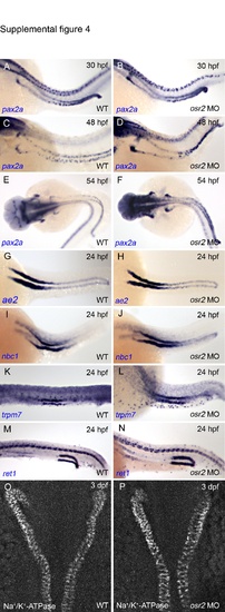
Pronephric kidney patterning and structure is normal in osr2 morphants. A–N: In situ hybridization of wild-type (A, C, E, G, I) and osr2 morphant embryos (B, D, F, H, J) using pronephric nephron markers. pax2a (A–F) is expressed in the entire pronephric nephron, ae2 (G and H) and nbc1 (I and J) are expressed in the proximal nephron, trpm7 (K, L) are expressed in the early distal segment and ret1 (M, N) is expressed in the distal segment of the nephron. Embryonic stages are indicated on the corresponding panels. O, P: a6F immunohistochemistry on wild-type (O) and osr2 morphant (P) showing normal expression of Na+/K+-ATPase in the pronephros. |
|
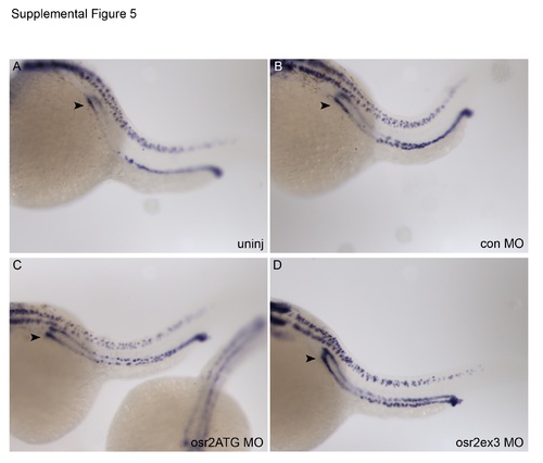
pax2a expression is unchanged in osr2 morphants. In situ hybridization of 24-hpf embryos using pax2a probe shows that wild-type uninjected controls (A) show roughly equivalent staining compared to control morphants (B). A slight increase in pax2a signal in translation-blocking (C) and splice-blocking (D) osr2 morphants may be due to greater probe penetration since pax2a signal was also more intense in the neural tube, which is not an osr2-expressing tissue. Arrowheads indicate anterior tubules where osr2 is normally expressed. |

