- Title
-
Zebrafish Foxi1 provides a neuronal ground state during inner ear induction preceding the Dlx3b/4b-regulated sensory lineage
- Authors
- Hans, S., Irmscher, A., and Brand, M.
- Source
- Full text @ Development
|
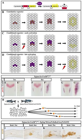
Pioneer tracking reveals that early OEPD pax2a-positive cells predominantly contribute to the anterior-ventral part of the otic vesicle, including the neurogenic region. (A) Removal of a loxP-flanked transcriptional STOP cassette (red) in the presence of Cre (purple) activates reporter expression (yellow). (B) In a common genetic lineage-tracing setup, Cre expression and reporter activation are directly linked by the constitutive promoter driving reporter expression. Consequently, the reporter is active in all Cre-positive cells even at later stages when Cre expression has vanished. (C) By contrast, use of the conditional temperature-inducible hsp70l promoter uncouples Cre expression and reporter activation. Early activation (red thunderbolt) restricts reporter expression to cells that experience recombination within a nascent Cre domain and allows fate mapping of these cells due to reporter protein persistence at later stages. (D) Late activation (red thunderbolt) of the conditional promoter reveals the fate of the entire Cre domain at later stages similar to a constitutive promoter. (E-G) CreERT2 (blue) recapitulates the dynamic, endogenous pax2a expression (red) during OEPD stages (3-, 4-, 6-somite) in transgenic Tg(pax2a:CreERT2)#31 embryos shown by two-color in situ hybridization. (E2-G2) High magnification views of the OEPD region of embryos shown in E-G, respectively. (H) Schematic of the experimental outline. The progeny of the indicated cross were exposed to tamoxifen (TAM) to elicit immediate Cre-mediated recombination as soon as CreERT2 is active. Subsequently, the offspring were divided into different groups, exposed to a single heat treatment at various developmental stages [3-, 6-, 9-, 14-somites (so)] and analyzed at 24 hpf. (I,J) EGFP-labeled cells are found in an anterior-ventral position within the otic vesicle after heat shock at the 3- or the 6-somite stage in Tg(pax2a:CreERT2)#31; Tg(hsp70l:loxP-DsRed-loxP-EGFP) double transgenic embryos. (K) Heat shock at the 9-somite stage expands the EGFP-positive domain without labeling posterior-dorsal positions within the otic vesicle. (L) EGFP-labeled cells can be detected throughout the otic vesicle after heat shock at placodal stages. E-G are dorsal views with anterior to the top at 3-, 4-, 5- and 12-somite stages. I-L are lateral views with anterior to the left at 24 hpf. Scale bars: in L, 90 μm for E-G; in L, 30 μm for E2-G2 in L, 40 μm for I-L. EXPRESSION / LABELING:
|
|
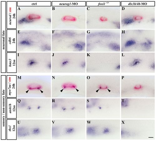
Foxi1 and Dlx3b/4b regulate the neuronal and sensory lineages of the inner ear. (A-X) Blue: Expression of neurod (A-D), cdh6 (E-H), hmx3 (I-L), myo7aa (M-P), atoh1b (Q-T) and tbx1 (U-X) in control (A,E,I,M,Q,U), neurog1-MO injected (B,F,J,N,R,V), foxi1 mutant (C,G,K,O,S,W) and dlx3b/4b-MO injected embryos (D,H,L,P,T,X). Red: Expression of stm reveals the size of the otic vesicle, which is reduced in foxi1 mutant and dlx3b/4b-MO injected embryos. A-H,M-P are lateral views with anterior to the left at 24 hpf. I-L,Q-X are dorsolateral views with anterior to the left at the 12-somite stage. Arrowheads indicate the position of the sensory patches. Scale bar: 40 μm. EXPRESSION / LABELING:
PHENOTYPE:
|
|
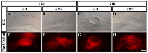
Fate mapping of OEPD Pax8-expressing cells in wild-type and b380 mutant embryos. (A-H) Live images of Pax8:DsRed in wild-type (A,C,E,G) and b380 mutant (B,D,F,H) embryos at the 12-somite stage (A,B,E,F) and 24 hpf (C,D,G,H), respectively. Lateral views with anterior to the left. Scale bar: 40 μm. EXPRESSION / LABELING:
PHENOTYPE:
|
|
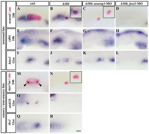
Persistent OEPD-dependent neurogenesis in Dlx3b/4b- and Sox9a-deficient b380 mutants. (A-R) Blue: Expression of neurod (A-D), cdh6 (E-H), hmx3 (I-L), myo7aa (M,N), atoh1b (O,P) and tbx1 (Q,R) in control (A,E,I,M,O,Q), b380 mutant (B,F,J,N,P,R), b380; neurog1-MO-injected (C,G,K) and b380; foxi1-MO-injected (D,H,L) embryos. Red: Expression of stm reveals the size of the otic vesicle which is reduced to a small epithelial ball in b380 mutant and b380; neurog1-MO-injected and entirely absent in b380; foxi1-MO-injected embryos. A-H,M,N are lateral views with anterior to the left at 24 hpf. I-L,O,R are dorsolateral views with anterior to the left at the 12-somite stage. Insets in B, C and N show higher magnifications of the remaining otic tissue. Arrowheads in M indicate the position of the sensory patches. Scale bar: 40 μm. EXPRESSION / LABELING:
PHENOTYPE:
|
|
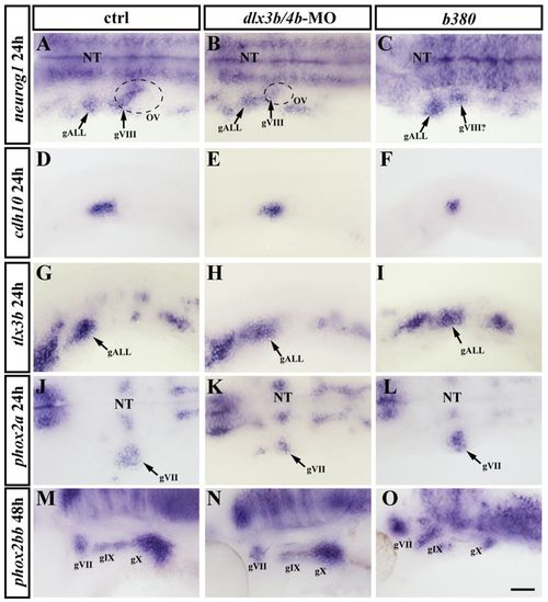
All OEPD-derived neuronal progenitors are present in Dlx3b/4b-depleted embryos and Dlx3b/4b- and Sox9a-deficient b380 mutants. (A-O) Expression of neurog1 (A-C), cdh10 (D-F), tlx3b (G-I), phox2a (J-L) and phox2bb (M-O) in control (A,D,G,J,M), Dlx3b/4b-depleted (B,E,H,K,N) and b380 mutant embryos (C,F,I,L,O). A-C,J-L are dorsal views with anterior to the left at 24 hpf. D-I,M-O are lateral views with anterior to the left at 24 hpf (D-I) and 48 hpf (M-O). gVII, geniculate ganglion; gVIII, statoacoustic ganglion; gIX, petrosal ganglion; gX nodose ganglion; NT, neural tube; OV, otic vesicle. Scale bar: 50 μm. |
|
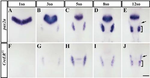
Expression of CreERT2 recapitulates the endogenous pax2a expression during OEPD development. (A-J) The temporal and spatial expression of CreERT2 (F-I) within the OEPD is identical to pax2a (A-D) in Tg(pax2a:CreERT2)#31 transgenic embryos. Subsequently, pax2a (E) is maintained exclusively in the otic placode (bracket) but is downregulated in non-incorporated cells anteriorly to it (arrow), whereas CreERT2 (J) is sustained in both regions. Dorsal views with anterior to the top at the 1-, 3-, 5-, 8- and 12-somite (so) stages, as indicated. Scale bar: 100 μm. |
|
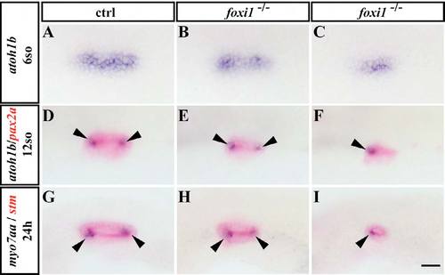
Hair cell formation is highly variably in foxi1 mutants, which is foreshadowed by atoh1b expression at OEPD stages. (A-I) Blue: Expression of atoh1b (A-F) and myo7aa (G-I) in control (A,D,G) and foxi1 mutant embryos (B,C,E,F,H,I). Red: Expression of pax2a and stm reveal size of the otic placode and vesicle, respectively. A-C are dorsal views with anterior to the top at the 3-somite stage. D-F are dorsolateral views with anterior to the left at the 12-somite stage. G-I are lateral views with anterior to the left at 24 hpf. Arrowheads indicate the position of the sensory patches. Scale bar: 35 μm. |
|
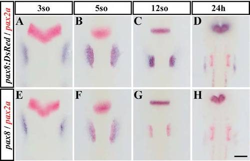
Expression of DsRed in a gene trap in which the coding sequence of DsRed is inserted into the pax8 locus, recapitulates the endogenous pax8 expression during otic development. (A-H) Blue: Expression of DsRed (A-D) and pax8 (E-H) in pax8nia03Gt transgenic embryos. Red: Expression of pax2a. Dorsal views with anterior to the top at the 3-, 5-, 12-somite (so) stage and 24 hpf, as indicated. Scale bar: 100 μm. |

