- Title
-
Analysis of her1 and her7 Mutants Reveals a Spatio Temporal Separation of the Somite Clock Module
- Authors
- Choorapoikayil, S., Willems, B., Ströhle, P., and Gajewski, M.
- Source
- Full text @ PLoS One
|
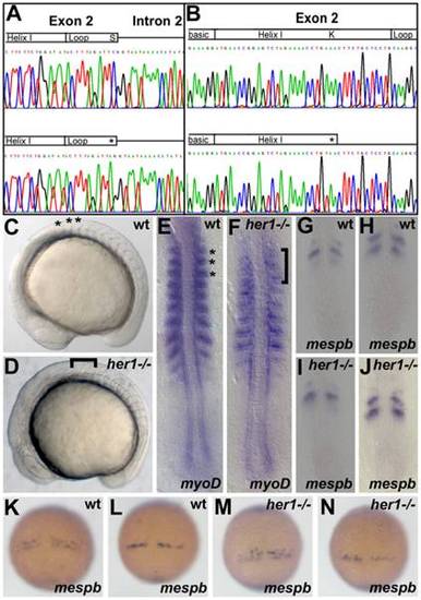
Her1 mutants exhibit defects in somitogenesis. Electropherogram of her1 (A) and her7 (B) amplicons in wild type (top), homozygous her1hu2124 (bottom, A) and her7hu2526 (bottom, B) mutant fish. Schematics above the sequences depict the exon- and intron-organization and the protein domains encoded by the exons. Point mutations are indicated by asterisks. (C, D) Brightfield pictures of wild type and her1 mutant embryos, lateral view, anterior to left. Compared to wild type embryos (C, asterisks), the first 3 somitic borders in the her1hu2124 mutant appear diffuse and partly disrupted (D, bracket). In situ analysis of myoD expression in wild types indicates characteristic half-segmental expression within the somites (E, asterisks indicate somites 1–3). myoD expression is diffuse in the first 3 somites of her1hu2124 embryos (F, bracket). (G, H) and (K, L) show half-segmental respectively r/c polarity wild type expression pattern of mespb at 10–12 somite stage and between 90% epiboly and bud stage, respectively. (I, J) and (M, N) represents mespb expression in the her1hu2124 mutant at 10–12 somite stage and between 90% epiboly and bud stage, respectively. Expression of mespb is disturbed in the her1hu2124 mutant between 90% epiboly and bud stage (M, N), when the anlagen of the first somites are pre-patterned. Compared to one or two stripes in the wild-type (K, L), mespb is expressed in a salt and pattern (M, N). mespb expression is unperturbed at 10–12 somite stage in her1hu2124 mutants (I, J). Dorsal views, anterior to top. EXPRESSION / LABELING:
PHENOTYPE:
|
|
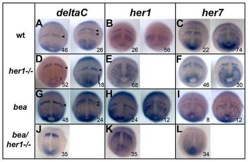
Expression analysis of segmentation genes in her1 hu212 and beatm98 mutants. In situ hybridisation analysis of segmentation clock genes deltaC, her1, and her7 in wild type (A–C) her1hu2124 mutants (D–F), bea tm98 mutants (G–I) and her1hu2124/bea tm98 double mutants (J–L) at 90% epiboly. Cyclic deltaC expression is disrupted in the anterior PSM of her1hu2124 mutants. Instead of one or two expression stripes as in the wild type (A, arrowheads) only one stripe of expression is observed (D, arrowhead). Expression domains in the posterior PSM display different sizes indicating unperturbed oscillation of deltaC in the tail bud of her1hu2124 mutants (D, bars). Cyclic expression of her1 is fully disrupted in the her1hu2124 mutant (E) when compared to wild type (B), whereas her7 expression remains oscillatory (compare C and F). Cyclic expression of all three genes is observed in beatm98 although some slight initial perturbation is observed (G-I). In her1hu2124/beatm98 double mutants, all three clock genes show fully disrupted expression patterns at 90% epiboly. Dorsal views, anterior to the top, number in each panel indicate cycling phases. EXPRESSION / LABELING:
|
|
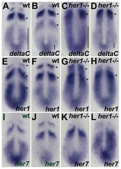
Expression analysis of the segmentation clock genes at 10–12 somite stage in her1hu2124 mutants. In situ hybridisation analysis of the segmentation clock genes deltaC, her1, and her7, in wild type embryos (A,B,E,F,I,J) her1hu2124 mutant (C,D,G,H,K,L) at the 10–12 somite stage. Two significantly different patterns are shown for each gene to indicate oscillatory expression. Expression of deltaC in her1hu2124 mutants at this developmental stage is identical to the 90% epiboly (see Fig. 2D), cyclic in the posterior PSM and disrupted expression in the anterior PSM (C, D) compared to wild type (A, B). Expression of her1 and her7 oscillates in the her1hu2124 mutant but on average one expression stripe is lacking (see asterisks in G, H and K, L, respectively) compared to the respective wild type expression domains (asterisks in E, F and I, J). Further, the patterns in the PSM of mutants appear stretched towards the anterior compared to wild type (see bars in A-D) suggesting that one expression wave is lacking. Dorsal view, anterior to the top. EXPRESSION / LABELING:
PHENOTYPE:
|
|
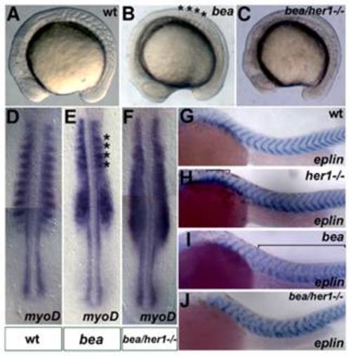
Analysis of the her1hu2124/bea tm98 double mutant phenotype. Brightfield images of wild type, beatm98 and her1hu2124/beatm98 mutant embryos at the 10–12 somite stage, lateral views, anterior to left. Compared to the wild type embryo (A), the somite borders posterior of the 4th somite are disrupted in the beatm98 mutant (B, asterisks indicate correctly formed somites). All somitic borders are disrupted in the her1hu2124/beatm98 double mutant (C). In situ hybridisation analysis of myoD expression at 10–12 somites (D–F), dorsal views, anterior to top. In line with the morphological phenotypes, half segmental myoD expression is disrupted posterior to the 4th somite in beatm98 (E, asterisks mark residual expression in somites 1–4) and along the entire body axis in her1hu2124/beatm98 double mutants (F) compared to wild type (D). In situ analysis of eplin expression at prim 6 stage (G–J) lateral views, anterior to left. eplin is expressed in v-shape at the somite borders in the wild-type (G). Disturbed eplin expression is observed in the first somites of the her1hu2124 mutant (H, bracket), posterior to the somite 4 in the beatm98 mutant (I, bracket) and in all somites in the double mutant situation (J). (A–F) 10–12 somite stage, (G–J) prim 6 stage. EXPRESSION / LABELING:
PHENOTYPE:
|
|
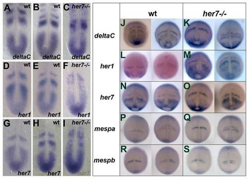
Expression analysis of segmentation genes in her7hu2526 mutant embryos. In situ hybridisation analysis of deltaC, her1 and her7 in wild type (A, B and D, E and G, H, respectively) and her7hu2526 mutants (C, F, I, respectively) at 10–12 somite stage and between 90% epiboly and bud stage (J, L, N for wild type expression patterns and K, M, O for respective expression patterns in the mutant embryos). Expression patterns of deltaC, her1 and her7 at 10–12 somite stage are disrupted in the mutant appear unperturbed between 90% epiboly and bud stage. Expression patterns of mespa and mespb are not affected in the her7hu2526 mutant between 90% epiboly and bud stage (Q and S, respectively) and similar to the wild type (P and R, respectively). EXPRESSION / LABELING:
|
|
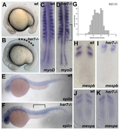
The role of Her7 during pre-pattering. Brightfield images of wild type and her7 mutant embryos at 16–18 somites (A, B). Compared to the wild type embryo (A), somite borders posterior to the 8th somite are disrupted in the her7hu2526 mutant (B, bracket). In situ hybridisation analysis of myoD expression (C, D), eplin (E, F), mespb (H, I) and mespb (J, K) in wild type and her7 mutants. Compared to half-segmental myoD expression in the wild type (C), myoD expression is disrupted posterior to the 8th somite in her7hu2526 mutants at 10–12 somites (D, bracket). In addition to the ALD at somite 8, her7hu2526 mutant larvae show a PLD at around somite 17 (F, bracket indicates area of defect). eplin expression posterior to the PLD appears V-shaped as in wild-type at prim 6 stage (compare E, F). (G) graph plotting the number of her7hu2526 embryos exhibiting defective somites (n = 56) as a function of their respective position along the a/p-axis of the animal. The obtained formula for the defect in the her7hu2526 mutant is 8 (+/3)17 (+/3) indicating that in some rare cases the defects seem to appear at both the ALD and the PLD with a slight variability. mespb expression in the wild type and the her7hu2526 mutant are shown in (H) and (I), respectively. (J) and (K) mespa expression in the wild type and her7hu2526 mutant, respectively. Expression of both genes is disrupted in the her7hu2526 mutant at 10–12 somite stage. (A, B, E, F) lateral view, anterior to the left; (C, D, H-K) dorsal view, anterior to the top. EXPRESSION / LABELING:
PHENOTYPE:
|

