- Title
-
Cabin1 expression suggests roles in neuronal development
- Authors
- Hammond, D.R., and Udvadia, A.J.
- Source
- Full text @ Dev. Dyn.
|
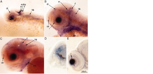
Cabin1 is expressed in the developing zebrafish nervous system. The developmental Cabin1 gene expression pattern as determined by mRNA in situ hybridization (ISH) in whole embryos is shown. A–C: Lateral views of the head at 24–72 hours postfertilization (hpf). D,E: Coronal sections through stained embryos at 72 hpf. A: At 24 hpf, expression is observed in the diencephalon (di), midbrain (m), midbrain–hindbrain boundary (m/hb), cerebellum (c), and hindbrain (hb). B: At 48 hpf, Cabin1 expression expands to the ganglion cell layer of the retina (r), and the olfactory epithelium (oe), and is also prominently expressed extraneuronally in the pharyngeal arches (pa). C,D: By 72 hpf, expression within the nervous system is still observed in the olfactory epithelium, cerebellum, and hindbrain, as well as in the diencephalon (C; in D, thalamus [th], preoptic area [po]). E: Expression in the retina at 72 hpf, has diminished to regions that correspond to regions where new neurons are generated. EXPRESSION / LABELING:
|
|
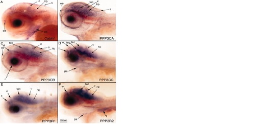
Cabin1 and calcineurin expression overlaps in the central nervous system (CNS). Calcineurin expression is widespread throughout the developing nervous system. Expression of Cabin1 coincides with one or more calcineurin genes in the olfactory epithelium (oe), cerebellum (c), and hindbrain (hb). A–F: Lateral views of the head at 72 hpf after whole embryo mRNA in situ hybridization (ISH) using probes for the following: Cabin1 (A) and all five calcineurin subunits: PPP3CA (B), PPP3CB (C), PPP3CC (D), PPP3R1 (E), PPP3R2 (F). Calcineurin expression at 24–72 hpf can been viewed in supplementary material (S1). di, diencephalon; e, epiphysis; l, liver; pa, pharyngeal arches; r, retina; s, skin; t, telencephalon; tec, tectum; teg, tegmentum. Some embryos have been manually deyolked. EXPRESSION / LABELING:
|
|
Cabin1 and MEF2 expression overlaps in the central nervous system (CNS). MEF2 isoforms exhibit distinct but overlapping gene expression patterns. A: At 48 hours postfertilization (hpf), MEF2A-Cb all have some overlap in the CNS with Cabin1 (A), which is most prominent in the midbrain (m), cerebellum (c), and hindbrain (hb). A–F: Lateral views of the head at 48 hpf after whole embryo mRNA ISH using probes for the following: Cabin1 (A), MEF2A (B), MEF2B (C), MEF2Ca (D), MEF2Cb (E), and MEF2D (F). MEF2 expression at 24–72 hpf can be viewed in the Supplementary Material (Supp. Fig. S2). di, diencephalon; fb, fin bud; h, heart: oe, olfactory epithelium; pa, pharyngeal arches; png, peripheral nerve ganglia; t, telecephalon. Some embryos have been manually deyolked. EXPRESSION / LABELING:
|
|
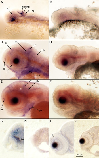
Cabin1 is expressed in the developing zebrafish nervous system. A–J: Embryos probed for Cabin1 sense control (B,D,F,H,J) contrast embryos probed with anti-sense Cabin1 (A,C,E,G,I) as determined by in situ hybridization (ISH). Lateral views of the head at 24–72 hours postfertilization (hpf; A–F) and coronal sections through stained embryos at 72 hpf (G–J). c, cerebellum; di, diencephalon; hb, hindbrain; m, midbrain; m/hb, midbrain/hindbrain boundary; oe, olfactory epithelium; pa, pharyngeal arches; po, preoptic area; r, retina; th, thalamus. EXPRESSION / LABELING:
|
|
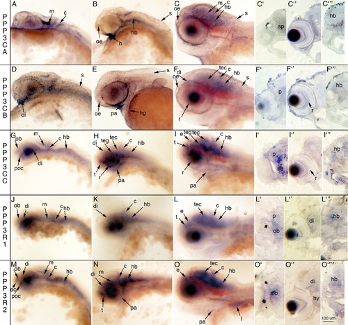
Calcineurin subunits are expressed in unique temporal and spatial patterns. All calcineurin subunits are expressed in the nervous system during development as determined by whole embryo in situ hybridization (ISH). A–O: Lateral views of the head at 24 hpf (A,D,G,J,M), 48 hpf (B,E,H,K,N), and 72 hpf (C,F,I,L,O) and coronal sections of stained embryos at 72 hours postfertilization (hpf; C2–C23 F2–F23 I2–I23 L2–L23 O2–O23). ac, anterior commissure; c, cerebellum; di, diencephalon; e, epiphysis; h, heart; hb, hindbrain; hg, hatching gland; hy, hypothalamus; l, liver; m, midbrain; ob, olfactory bulb; oe, olfactory epithelium; p, pallium; pa, pharyngeal arches; poc, post optic commissure; r, retina; s, skin; sp, subpallium; t, telencephalon; tec, tectum; teg, tegmentum. Some embryos have been manually deyolked. EXPRESSION / LABELING:
|
|
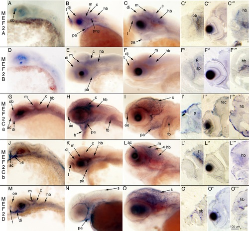
MEF2 isoforms are expressed in distinct temporal and spatial expression patterns in the developing CNS. All MEF2 isoforms are expressed in the nervous system during development as determined by whole embryo ISH. Lateral views of the head at 24 hpf (A, D, G, J, M), 48 hpf (B, E, H, K, N), and 72 hpf (C, F, I, L, O) and coronal sections of stained embryos at 48 hpf (I2–I23) and 72 hpf (C2–C23 F2–F23 L2–L23 O2–O23). ac, anterior commissure; c, cerebellum; di, diencephalon; fb, finbud; h, heart; hb, hindbrain; m, midbrain, ob, olfactory bulb; oe, olfactory epithelium; p, pallium; pa, pharyngeal arches; png, peripheral nerve ganglia; s, skin; sp, subpallium; t, telencephalon; tec, tectum. Some embryos have been manually deyolked. EXPRESSION / LABELING:
|

