- Title
-
Adaptive autoimmunity and foxp3-based immunoregulation in zebrafish
- Authors
- Quintana, F.J., Iglesias, A.H., Farez, M.F., Caccamo, M., Burns, E.J., Kassam, N., Oukka, M., and Weiner, H.L.
- Source
- Full text @ PLoS One
|
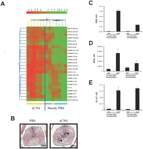
Adaptive autoimmunity in zebrafish. (A) Heatmap depicting the autoantibody response to myelin antigens on day 28 after immunization with zCNS or PBS in CFA. Each column represents a serum sample, color-coded at the bottom to indicate whether it corresponds to a zCNS or a control immunized sample. Only significantly up-regulated antibody reactivities are shown (n = 8, t-test FDR <0.05), according to the colorimetric scale on the right. (B-E) Zebrafish were immunized with zCNS or PBS in CFA and 14 or 28 days later the expression of CD3, IL-17 and IFNγ in brain was measured by real time PCR (mean + s.d. of triplicates) (B-D) or analyzed histologically for the presence of cell infiltrates (E). Two independent experiments produced similar results. |
|
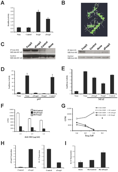
zFoxp3 is a functional homologue of mammalian Foxp3. (A) Constructs coding for His-labeled zFoxp3 and Renilla-labeled Foxp3 were co-transfected into 293T cells. 24 h later the cells were lysed, zFoxp3 was pulled-down with Ni-Agarose and the renilla luciferase activity in the pellet was quantified. The results are normalized for the total amount of luciferase before precipitation (mean + s.d. of triplicates). Three independent experiments produced similar results. (B) Structure of the forkhead domain of zFoxp3 obtained by homology modeling, based on the structure of the crystallized forkhead domain of Foxp1. (C) 293T cells were co-transfected with His-tagged zFoxp3, Foxp3 and NF-κB or HA-flagged NFAT and 24 hr later the cells were lyzed and immunoprecipitated with antibodies to His antibodies. The precipitates were resolved by PAGE-SDS and detected by western blot with antibodies to NF-κB or HA antibodies. Three independent experiments produced similar results. (D, E) 293T cells were co-transfected with reporter constructs coding for luciferase under the control of a NF-κB or NFAT responsive promoters, and p65 NF-κB or NFAT in the presence of vectors coding for zFoxp3, Foxp3 or control (empty vector). Luciferase activity was normalized to the renilla activity of a co-transfected control (mean + s.d. of triplicates). Four independent experiments produced similar results. (F) MACS-purified CD4+CD25- T-cells were transduced with a bicistronic retrovirus coding for GFP and zFoxp3, Foxp3 or an empty control retrovirus, and the GFP+ population was analyzed for its proliferation upon activation with plate bound antibodies to CD3 (mean cpm or pg/ml + s.d. in triplicate wells) and (G) its suppressive activity on the proliferation and IL-2 and IFNγ secretion of mouse CD4+CD25- T-cells activated with plate-bound antibodies to CD3 (mean cpm or pg/ml + s.d. in triplicate wells). Two independent experiments produced similar results. (H) Fertilized zebrafish eggs were microinjected with a plasmid coding for zFoxp3 or an empty plasmid, and zFoxp3 and IL-17 expression were measured from 6 days old embryos by real time PCR. Two independent experiments produced similar results. (I) Fertilized zebrafish eggs were microinjected with a morpholino oligonucleotides designed to interfere with the translation of zFoxP3 (Mo-zFoxp3) or a 5 bases mismatch control oligonucleotide and IL-17 expression was measured in 5 days old embryos by real time PCR. Two independent experiments produced similar results. EXPRESSION / LABELING:
|
|
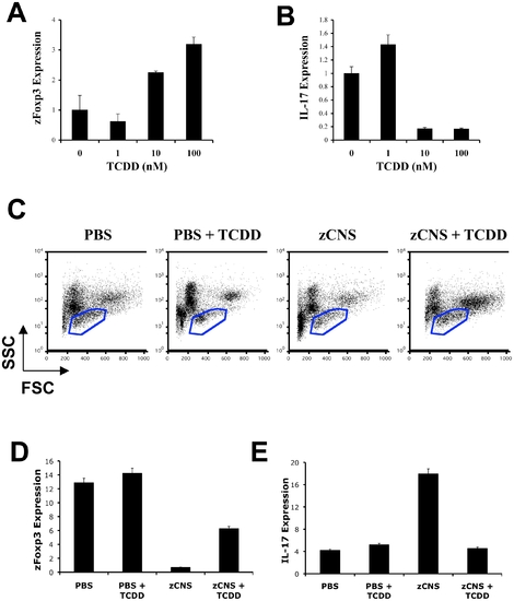
AHR controls zFoxp3 expression. (A,B) TCDD was added to the water of three-day post-fertilization zebrafish embryos, and 72 h later zFoxp3 (A) and IL-17 (B) expression were determined by real time PCR (mean + s.d. of triplicates normalized to GAPDH expression). Two independent experiments produced similar results. (C) Fourteen days after immunization, kidney cells from PBS or zCNS immunized zebrafish, or TCDD-treated zCNS immunized zebrafish were analyzed by FACS and cells in the lymphocyte fraction (blue gate) were sorted. (D–E) Expression of zFoxp3 and IL-17 measured by real-time PCR in FACS-sorted lymphocytes. Two independent experiments produced similar results. EXPRESSION / LABELING:
|


