- Title
-
Bucky Ball Organizes Germ Plasm Assembly in Zebrafish
- Authors
- Bontems, F., Stein, A., Marlow, F., Lyautey, J., Gupta, T., Mullins, M.C., and Dosch, R.
- Source
- Full text @ Curr. Biol.
|
Germ Plasm Formation in Early Bucky Ball Oocytes and Evolutionary Conservation of the Buc Protein (A–F) Fluorescent whole-mount in situ hybridization of early Ib oocytes showing mRNAs (red) of cyclinB (A and D), nanos (B and E), and vg1RBP (C and F) in wild-type (A–C) and buc p106re mutant oocytes (D–F). Note that the localization of nanos and vg1RBP mRNAs in the Balbiani body is lost in mutants (E and F), but the cyclinB and vg1RBP mRNAs still show polarized localization (D and F). Animal pole to the top. Scale bar represents 25 μm. All mutant images are of the bucp106re allele. (G) Unrooted star diagram displaying the phylogenetic conservation of Bucky ball proteins among vertebrates. The scale indicates the number of substitutions per amino acid residue. (H) Alignment of Buc N terminus in 12 vertebrate species (zebrafish amino acids 23–136). The sequence homology was calculated with Kalign [45], which revealed a conserved peptide (zebrafish amino acids 23–38, red box). (I) Alignment of conserved peptide (red box in H) with T-Coffee [46]. The color code below the alignment illustrates the level of conservation (cons.) from BAD (blue) to GOOD (red). Note that, with evolutionary distance, the sequence similarity decreases. Gene-ID from NCBI or Ensembl-genome databases: Zebrafish, Danio rerio: NCBI-locus 560382; Medaka, Oryzias latipes: UTOLAPRE05100115054; Spotted pufferfish, Tetraodon nigroviridis: GSTENT00016539001; Tiger pufferfish, Takifugu rubripes: SINFRUT00000183089; Clawed frog, Xenopus tropicalis: ENSXETT00000045658; Chicken, Gallus gallus: ENSGALT00000019911; Platypus, Ornithorhynchus anatinus: GENSCAN00000155561; Opossum, Monodelphis domestica: GENSCAN00000062731; Cow, Bos taurus: GENSCAN00000042786, GENSCAN00000042790; Pig, Sus scrofa: Ssc.46160, Ssc.28451; Dog, Canis familiaris: GENSCAN00000091809, GENSCAN00000058820; Man, Homo sapiens: EU128483, EU128484 |
|
Expression Analysis of buc mRNA (A–D) Real-time PCR analysis of buc and vasa mRNA relative to expression in the one-cell embryo during oogenesis (A), embryogenesis (B), and in sexually mature adults (C). (D) Real-time PCR comparing the levels of buc, dazl, and vasa mRNA in wild-type (+/-) and bucp106re mutant ovaries (-/-). (E–L) Fluorescent whole-mount in situ hybridization of buc (red in all pictures) during stage I (E–H) and stage III (I–L) of oogenesis. Animal to the top in all pictures. (F and J) Double in situ hybridizations showing colocalization of buc and the vegetal dazl mRNA (green) at stage I (F), but not at stage III (J). (G and K) Double in situ hybridizations showing colocalization of buc and foxH1 mRNA (green) at stage I (G) and at stage III (K). (H and L) Animal localization of buc mRNA at stage I (H), but not after 3-fold longer exposure at stage III in bucp106re oocytes (L). Scale bars represent 25 μm (E–H) and 50 μm (I–L). |
|
buc mRNA Translation Is Required to Assemble Germ Plasm, and Buc-GFP Localizes to the Balbiani Body (A–E) Buc is required in the oocyte for germ plasm localization. Wild-type (A and C) and bucp106re mutant oocytes (B and D) after microinjection of wild-type (A and B) or mutant bucp106re mRNA (C and D) into stage I zebrafish oocytes. (E) Quantification of dazl mRNA localization from three independent experiments in WT oocytes, uninjected (75% ± 6.0%, n = 63), injected with WT buc (84% ± 14%, n = 67), or mutant bucp106re mRNA (83% ± 12%, n = 47) compared to bucp106re mutant oocytes, uninjected (6% ± 3.9%, n = 122), injected with WT buc (26% ± 5.1%, n = 103), or mutant bucp106re mRNA (7% ± 6.4%, n = 26). Note the rescue of dazl mRNA localization in mutant oocytes after injection of wild-type buc mRNA (B and E). Error bars represent SD. ***p = 0.0008. (F–I) Buc needs to be translated to assemble germ plasm. bucp106re mutant stage I oocytes injected with mutant bucp106re (F and H) or WT buc mRNA (G and I) with control (+ctrl-MO) (F and G) or translation inhibition morpholino (+buc-MO) (H, I), which inhibited translation of Buc-GFP fusion mRNA and, hence, GFP-fluorescence efficiently in embryos (Figure S5). (A–D and F–I) Whole-mount in situ hybridization of dazl mRNA (blue). Scale bars: 25 μm. (J) Quantification of dazl mRNA localization in mutant oocytes injected with mutant bucp106re mRNA plus ctrl-MO (9%, n = 11) or plus buc-MO (10%, n = 21) and WT mRNA plus ctrl-MO (25%, n = 20) or plus buc-MO (11%, n = 27). Note that the rescue of dazl mRNA localization is blocked by a morpholino-inhibiting buc mRNA translation (I and J). (K–M) Buc localizes to the Balbiani body. Living WT oocytes injected with mRNA encoding a Buc-GFP fusion (green) at mid (K) and late-stage Ib (L), as well as stage II (M) of oogenesis. Scale bars represent 25 μm. (N) Model of the role of Buc in early-stage I oocytes. Buc inhibits the premature localization of foxH1, vg1RBP, and buc mRNA at the animal pole (A) and concomitantly recruits the germ plasm RNAs dazl, nanos, and vasa in the Balbiani body at the vegetal pole (V). |
|
Buc Colocalizes with Germ Plasm and Induces the Formation of Germ Cells during Embryogenesis (A and B) Buc-GFP localizes to the germ plasm in eight-cell embryos. One-cell embryos were injected with 200 pg mRNA-encoding Buc-GFP (green) with (A) or without 3′UTR (B). Note the localization of GFP at four cleavage furrows in living eight-cell embryos. Animal view. Scale bars represent 200 μm. (C and D) buc mRNA is throughout the blastodisc during embryogenesis. Whole-mount in situ hybridization showing buc mRNA in the blastodisc (blue) without specific localization to the germ plasm at the cleavage furrow at four-cell (C) and blastula stages (D) (high stage). Animal view (C), lateral view, animal to the top (D). Scale bars represent 200 μm. (E–J) Overexpression of Buc in one-cell embryos induces germ cell formation. Dorsal view of living 13 somite stage embryos (15.5 hpf), anterior to the left (E and F). Germ cells are labeled green fluorescent after coinjection with 100 pg GFP-nanos-3′UTR mRNA. In control embryos, germ cells accumulate in the bilateral gonad anlagen (E), whereas in buc-injected embryos, extragonadal germ cells are visible (F). Animal view of oblong stage embryos (3.5 hpf) after whole-mount in situ hybridization with vasa mRNA (blue) injected with 300 pg GFP (G) or buc mRNA at the one-cell stage (H). Note additional vasa-positive cells in buc-injected embryos (85.0% ± 11.5%; n = 343) compared to control injections (5.7% ± 8.7%; n = 280; ***p = 3.4 x 10⁻¹²). Scale bars represent 200 μm (E–H). (I and J) Real-time PCR analysis of the germ plasm RNAs nanos, vasa, and dazl after control and buc mRNA injection analyzed at oblong stage (3.5 hpf; [I]) and 18 somites stage (18 hpf; [J]). Error bars represent SD (dazl *p = 0.028; vasa *p = 0.041; Table S3). (K and L) Experimental scheme for germ cell formation assay. (K) The 16-cell embryos (animal view) were either injected into one corner blastomere (green arrowhead) or into a middle blastomere (blue arrowhead; positive control) containing essential germ plasm (red ovals). (L) Embryos were examined between the 13 and 18 somite stage for the formation of additional germ cells (green dots) in addition to the endogenous germ cells (red dots; not visible in the experiment). Oblique dorsal view, anterior to the left. (M and N) Live 15 somite stage embryos, similar view as in panel (L), after injection of 100 pg GFP-nanos 3′-UTR mRNA into a corner blastomere (M) or after coinjection of 170 pg buc mRNA into the corner blastomere (N) with 12 ± 5.4 (n = 21) fluorescent cells per embryo. Scale bars represent 200 μm. (O and P) Buc-induced germ cells express vasa mRNA. Injection of GFP-nanos 3′-UTR into a corner blastomere of a 16-cell embryo does not label germ cells after vasa mRNA in situ hybridization at the 18 somite stage (black; [O]), whereas coinjection of buc mRNA generates additional vasa-positive cells also expressing GFP (green, white arrowheads; [P]). Dorsal view, anterior to the left. Scale bars represent 200 μm. |
|
Egg Polarity and Germ Plasm Formation in Early Bucky Ball Oocytes |
|
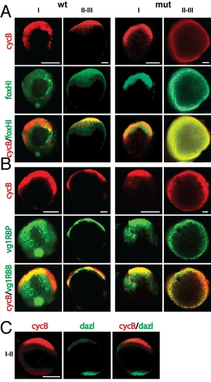
cyclinB, foxH1, and vg1RBP mRNA Localization in Wild-Type and buc Mutants |
|
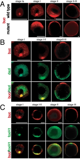
buc, dazl, and foxH1 mRNA Localization during Oogenesis |
|
Buc-Specific Morpholinos Inhibit Translation of buc-GFP Fusion mRNA |
|
Buc-GFP Overexpression Generates Additional Fluorescent Spots |


