- Title
-
A screen for genetic defects of the zebrafish ear
- Authors
- Schibler, A., and Malicki, J.
- Source
- Full text @ Mech. Dev.
|
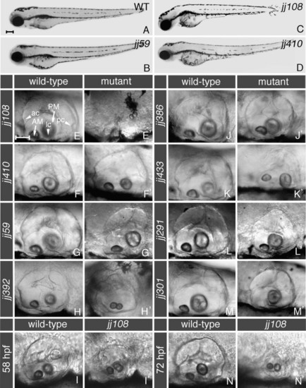
External phenotypes of ear morphology mutants. Panels (A-D) display lateral views of whole larvae at 72 hpf. A wild-type larva (A) is compared to mutant animals at the same stage (B-D). Note that the ear abnormalities of hako mimijj59 (B), mikre uchojj108 (C), and ale uchojj410 (D) mutants appear before other defects, indicating that they do not arise as a secondary consequence of a gross degeneration in the entire embryo. (E-N′) Lateral views of ear morphology in mutant animals, compared to their wild-type siblings. One of the most severe phenotypes is displayed by mikre uchojj108 mutants. Pictured at 144 hpf, the mkojj108 otic vesicle is severely reduced in size and malformed (E′, compare to E). mikre ucho mutant animals form two small otoliths that appear to be fused and their ears gradually decrease in size during larval development, so that they are smaller at 72 hpf (N′) compared to 58 hpf (I′). At 120 hpf, the ale uchojj410 mutant ear (F′) appears less differentiated compared to that of a wild-type sibling (F). Shown at 144 hpf, hako mimijj59 mutant ears display a shortened anterior-posterior axis (G′ compare to G). At 72 hpf, the smujj392 mutant ear is reduced in size in comparison to the wild type (H′ compare to H). pata nopojj386 (J′) mutant ears, pictured at 144 hpf, appear slightly smaller and more symmetric when compared to wild-type siblings (J). The gapajj433 mutant ear appears flat at 144 hpf (K′). Pictured at 144 hpf, the frostbite291 mutant ear is smaller and abnormally pointed on the dorsal side (L′ compare to L). The tone deafjj301 ear is smaller and features a single otolith, at 152 hpf (M′, compare to M). ac, anterior crista; lc, lateral crista; pc, posterior crista; AM, anterior macula; PM, posterior macula. In all panels, anterior is to the left and dorsal is up. Larvae were photographed using DIC optics. See Table 1 for a further description of phenotypes. Scale bar in (A) applies to panels (A-D), and equals 200 μm. Scale bar in (E) applies to the remaining panels, and equals 50 μm. |
|
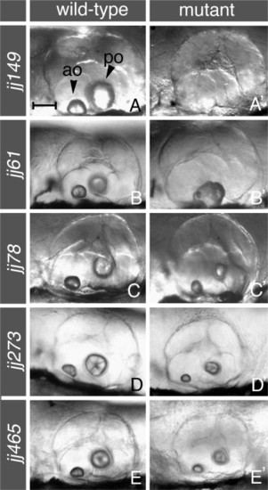
External phenotypes of otolith mutants. Lateral views of the ear in mutant animals (right column), compared to their wild-type siblings at the same stage (left column). At 144 hpf, no contentjj149 mutant embryos (A′) lack otoliths entirely. At the same stage, the rough edgesjj61 mutation (B′) usually results in one misshapen otolith characterized by a rough appearance, while teeny rocksjj78 (at 144 hpf), pebblesjj273 (at 120 hpf), and condensedjj465 (at 154 hpf) mutants (panels C′, D′, and E′ respectively) feature otoliths reduced in size when compared to their counterparts in wild-type siblings (C–E). Arrowheads in (A) indicate anterior (ao) and posterior (po) otoliths. In all panels, anterior is to the left and dorsal is up. Larvae were photographed using DIC optics. See Table 1 for a further description of phenotypes. Scale bar in (A) applies to all panels, and equals 50 μm. |
|
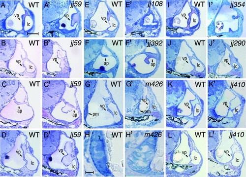
Histological analysis of morphology mutants. Transverse plastic section were prepared through ears of larval zebrafish. The first, the third and the fifth column from the left display sections of wild-type animals, while the second, the fourth, and the sixth column display sections of their mutant siblings at corresponding stages. The hako mimijj59 mutant phenotype is characterized by a thickening of inner ear pillars at 72 hpf (A′ compare to the wild type in A). By 120 hpf (B′), the ventral pillar of the kmijj59 ear frequently expands, blocking the lumen of the lateral canal (compare to the wild type in B). Panel (C′) displays a malformed kmijj59 anterior pillar characterized by an abnormal outgrowth at 120 hpf (compare to C). Another example of the kmijj59 mutant, featuring a bulb-like outgrowth instead of the ventral pillar is shown in (D′). The ear of mikre uchojj108 is strongly reduced in size at 72 hpf and its walls are thickened (E′, compare to E). Also at 72 hpf, smojj392 mutants (F′) feature a malformation of anterior pillar shape and size. At 144 hpf, antm426 mutant embryos (G′), display a reduced otic vesicle that is almost completely filled by the otolith. Enlargements of wild-type and antm426 mutant posterior maculae are shown in (H) and (H′), respectively. Note the malformation of the posterior macula in the antm426 mutant. Panel (I′) displays the pata nopojj354 mutant ear. At 72 hpf, its ventral pillar is incompletely formed. The ear of frostbitejj290 mutant is reduced at 144 hpf (J′, compare to J). In, ale uchojj410, epithelial protrusions that form the ventral pillar are thickened and incompletely formed at 72 hpf. (K′). By 120 hpf, however, ale uchojj410 ventral pillar displays a largely normal shape (L′). The following structures of the ear are indicated with arrows: ap, anterior pillar; vp, ventral pillar; pm, posterior macula. “lc” indicates lateral canal. In all panels, dorsal is up and lateral is right. Scale bar in (H) equals 10 μm, and applies to panels (H) and (H′). Scale bar in (A) equals 50 μm, and applies to all remaining panels. PHENOTYPE:
|
|
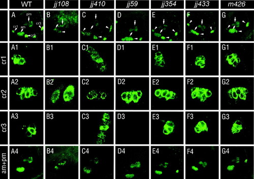
Hair cell distribution in mutants of ear morphology. Hair cells were evaluated by HCS-1 antibody staining at 5 dpf. Images of whole ears in wild-type and mutant animals are shown in the top row. Panels below, rows labeled cr1, cr2, and cr3, represent enlargements of anterior, lateral, and posterior cristae, respectively. The bottom row, labeled am + pm, shows the anterior and the posterior macula. The most severe phenotypes are observed in mikre uchojj108, ale uchojj410, and hako mimijj59 mutant animals. In mikre uchojj108, only a rudiment of one crista appears to be present (B2) and both maculae are severely misshapen (B4). A subpopulation of ale uchojj410 mutants exhibit disorganized and expanded cristae (C3). hako mimijj59 mutants lack both anterior and posterior cristae but retain the wild-type appearance of maculae (D4) and the lateral crista (D2). The remaining mutants do not display obvious defects of sensory patch morphology or positioning. In panels (A–G), cristae are indicated with arrowheads and maculae with arrows. The following structures are labeled in (A): cr1, anterior crista; cr2, lateral crista; cr3, posterior crista; am, anterior macula; and pm, posterior macula. In all panels, dorsal is up and anterior is left. PHENOTYPE:
|
|
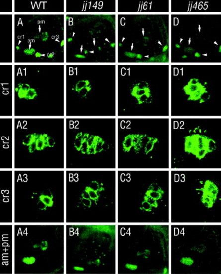
Hair cell morphology and distribution in otolith mutants. Hair cells were evaluated by HCS-1 antibody staining at 5 dpf. Images of whole ears in wild-type and mutant animals are shown in the top row. Panels below, in the rows labeled cr1, cr2, and cr3, show enlargements of anterior, lateral, and posterior cristae, respectively. The bottom row, labeled am + pm, shows the anterior and the posterior macula. No otolith mutants show obvious defects of hair cell distribution. In (A–D), cristae are indicated with arrowheads, and maculae with arrows. The following structures are labeled in (A): cr1, anterior crista; cr2, lateral crista; cr3, posterior crista; am, anterior macula; and pm, posterior macula. In all panels, dorsal is up and anterior is left. PHENOTYPE:
|
|
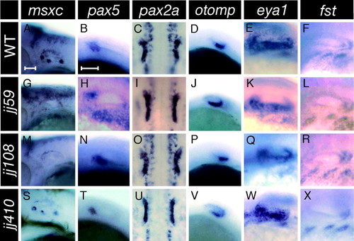
Early patterning of hako mimijj59, mikre uchojj108, and ale uchojj410 mutant ears. The expression of the anterior marker, pax5 (B, H, N, T), does not show any differences between wild-type and mutants. The same is true for pax2a (C, I, O, U), otomp (D, J, P, V), eya1 (E, K, Q, W), and follistatin (fst) (F, L, R, X). The msxc probe (A, G, M, S) is a marker of cristae. Its expression is absent in both mikre uchojj108 (M), and hako mimijj59 (G) mutants, but remains wild-type in ale uchojj410 (S). pax5, pax2a, otomp, and eya1 expression patterns were evaluated at 24 hpf, the fst expression at 36 hpf, and msxc at 48 hpf. In the pax2a column, expression is shown from the dorsal side. In all other columns, anterior is left and dorsal is up. Images of mxsc and otomp are at the same magnification. Likewise, the images of pax5, pax2a, eya1, and fst were collected at the same magnification. Scale bars equal 50 &mum. |

Unillustrated author statements |
Reprinted from Mechanisms of Development, 124(7-8), Schibler, A., and Malicki, J., A screen for genetic defects of the zebrafish ear, 592-604, Copyright (2007) with permission from Elsevier. Full text @ Mech. Dev.

