- Title
-
Osteocalcin and matrix Gla protein in zebrafish (Danio rerio) and Senegal sole (Solea senegalensis): Comparative gene and protein expression during larval development through adulthood
- Authors
- Gavaia, P.J., Simes, D.C., Ortiz-Delgado, J.B., Viegas, C.S., Pinto, J.P., Kelsh, R.N., Sarasquete, M.C., and Cancela, M.L.
- Source
- Full text @ Gene Expr. Patterns
|
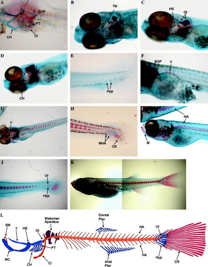
Time-course of skeletal development in zebrafish using Alcian blue-Alizarin red double staining. Whole mount double staining of the skeleton was used to follow ontogenic development of cartilaginous and calcified structures. (A) 96 hpf zebrafish larvae head skeleton presenting calcified pharyngeal teeth (PT), cleithrum (Cl), and basioccipital articulatory process (BOP) while other structures like Meckel′s cartilage (MC) and ceratohyal (CH) remain as cartilage (100x); (B) 5 dpf zebrafish larva presenting calcification on the opercula (Op) (40x); (C) beginning of the calcification of the hyosymplectic (HS) in a 6 dpf larva (100x); (D) Beginning of the calcification of the ceratohyal (CH) in a 8 dpf zebrafish (100x); (E) First hypurals (Hyp) developing at 8 dpf (100x); (F) The formation of the first vertebrae (V) is observed at 9 dpf (100x); (G) Calcification of the trabeculae (T) is observed at 11 dpf, vertebrae continue to form (in an anterior-posterior direction) towards the posterior end of the notochord (40x); (H) Caudal hypuralia acquires final number of structures with modified hemal arches (MHA) and caudal fin rays (CR) appearing at 12 dpf (100x); (I) Formation of the first neural arches (NA) is observed dorsally in the anterior vertebrae at 14 dpf; note that the mandibular is already calcified (M) (40x); (J) beginning of calcification of the hypurals (Hyp) under the urostile (Ur) at 14 dpf (40x); (K) composite image of a 19 dpf D. rerio with most skeletal structures formed and calcified; (L) schematic representation of the major zebrafish skeletal elements focused on this study. (SM) Supra-mandibular, (PT) pharyngeal teeth, (Ot) otolith, and (HA) hemal arches. |
|
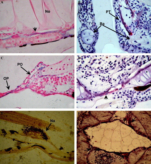
Alkaline phosphatase and TRAP activity in the developing zebrafish (A-D) and sole (E-F) skeleton. (A) Alkaline phosphatase activity (in blue) detected in the forming vertebra (arrow) surrounding the notochord (No) of a zebrafish larva at 13 dpf (1000x). (B) Tartrate resistant acid phosphatase (TRAP) activity (in red) is detected in developing, partially calcified, branchial arches (BA) and pharyngeal teeth (PT) of a zebrafish at 17 dpf (1000x). (C) Alkaline phosphatase activity in the calcifying preopercular cartilage (PO) and in the intramembranous opercular bone (OP) (1000x) in zebrafish larvae at 17 dpf. (D) TRAP activity in the intramembranously forming skull bones (1000x) of zebrafish at 26 dpf. A cell surrounded by TRAP activity is located in a resorption site (arrow). (E) Alkaline phosphatase activity (blue) is detected in 17 dpf Senegal sole in the forming neural arches (1000x). (F) TRAP activity (red) is detected surrounding the vertebra, arches, and spines. EXPRESSION / LABELING:
|
|
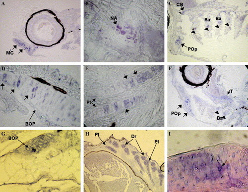
In situ localization of zebrafish and sole bgp mRNA. bgp gene expression was detected by in situ hybridization in sections of zebrafish (A–G) and sole (G–I) larvae collected at different ages and developmental stages. (A) bgp expression in the calcifying Meckel’s cartilage (arrows) of an 11 dpf larva (200×). (B) Detection of bgp expression in a neural arch (arrowhead) at the beginning of formation at 11 dpf (1000×). (C) At 13 dpf, bgp expression is detected in the branchial arches (BA) (arrowheads), ceratobranchial (CB), and calcifying preopercular bones (Pop) (arrow) (100×). (D) At 20 dpf, bgp expression is detected in chondrocytes of the BOP (arrows) (1000×). (E) Expression is visible in the mineralizing internal fin support skeleton, in this case the pterigophores (Pt) of the dorsal fin of a 24 dpf zebrafish (1000×). (F) At 24 dpf, bgp expression is strongly detected in the calcifying preopercular bones (arrows), branchial arches (arrowheads), and trabeculae (small arrow) (100×). (G) bgp expression in the first vertebra forming over the notochord (arrow), just posterior to the BOP at 15 dpf (400×). (H) Expression in the dorsal pterigophores (Pt) and distal radials (Dr) of a 20 dpf larvae (200×). (I) Expression in the dorsal pterigophores (Pt) and distal radials (Dr) of a 20 dpf larva (200×). (I) Osteoblasts (arrow) expressing bgp in the mandibula of a 56 DAH juvenile sole (1000×). For other abbreviations, see Fig. 1. EXPRESSION / LABELING:
|
|
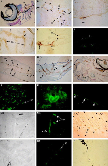
Immunohistochemical and immunofluorescent detection of Bgp accumulation at different developmental stages. (A–K) Bgp in zebrafish. (A) Accumulation of Bgp in the teeth of branchial arch 5 (Ba) and otolith (Ot) of a 8 dpf larva (200×); counterstaining with toluidine blue. (9B) Accumulation of Bgp in the teeth of branchial arch 5, otolith and cleithrum of a 9 dpf larva (400×). (C) Accumulation of Bgp in Meckel’s cartilage of a 13 dpf larva (1000×). (D) Accumulation of Bgp is first detected in calcifying vertebra and hemal arches at 13 dpf (1000×). (E) Accumulation of Bgp in the calcifying pectoral fin (F) and coraco–scapular complex (Co) at 20 dpf (400×). Notice the accumulation at the periphery of the coracoid undergoing perichondral mineralization. (F) Immunofluorescent detection of Bgp accumulation in the mineralizing urostile at 20 dpf (400×). (G) Accumulation of Bgp in the vertebral column (V), neural arches (Na), neural spines (Ns), and hemal arches (Ha) at 20 dpf (200×). (H) Bgp accumulation in the vertebral column, hemal arches, and in the mineralizing matrix of hypural plates (Hy) of the caudal fin and in the mineralized fin rays of a 31 dpf juvenile (100×). (I) Accumulation of Bgp in a juvenile is detected in all the bones and calcifying cartilages of the head region, e.g., supramaxillary (arrow) and associated bones, the ethmoid plate (arrowhead), and the supra orbital cartilage that surrounds the eye (100×). (J) Accumulation of Bgp is widely detected by immunofluorescence in the branchial arches of a juvenile (200×). (K) Bgp accumulation in epithelial cells from some renal tubules of the kidney in juveniles (400×). (L–P) Bgp in sole. L-Immunofluorescent detection of Bgp in the forming vertebra (arrows) surrounding the notochord of a 15 dpf larvae (400×). (M) Detection of Bgp accumulation in the vertebra and parapophysis (Pp) at 25 dpf (M1 bright field and M2 dark field) (250×). (N) Accumulation of Bgp in the calcifying distal radials (Dr) at 25 dpf. (O0 Bgp is strongly detected in the calcified branchial arches of a 41 dpf juvenile (O1 bright field and O2 dark field) (250×). (P) Control immunohistochemical detection with pre-immune serum lacks staining, shown here in cranial bones of a 20 dpf larvae. EXPRESSION / LABELING:
|
|
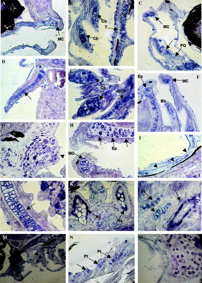
In situ localization of zebrafish and sole mgp mRNA. Sites of mgp gene expression were detected by in situ hybridization in sections of zebrafish (A–L) and sole larvae (M–O). (A) mgp expression in the ethmoid plate of a 96 hpf larva (200×). (B) Detection of mgp expression in chondrocytes from the trabecular cartilage (T) and ceratobranchial arches from a 9 dpf larva (Cb) (1000×). (C) mgp expression in Meckel’s cartilage (Mc) and palato quadrate (PQ) in a 9 dpf larva (100×). (D) Cartilage from the pectoral fin of a 10 dpf larva showing mgp expression located within the chondrocytes (arrow). Note the absence of signal in the cleithrum (asterisk) (1000×). (E) In 13 dpf larvae, mgp expression in the ceratobranchials (Cb) and in the BOP (1000×). (F) At 13 dpf, mgp expression was also evident in the hypertrophic chondrocytes from Meckel’s cartilage (Mc), ethmoid plate (Ep), and in chondrocytes from the basibranchial cartilage (Bb) (200×). (G) mgp expression in a 16 dpf larva, showing expression in chondrocytes from the BOP (arrowhead) (1000×). (H) At 16 dpf, mgp expression is also detected in hypertrophic chondrocytes of Meckel’s cartilage (Mc) and in chondrocytes from the ethmoid plate (Ep) (1000×). (I) 17 dpf larva showing mgp expression in chondrocytes from the optic capsules (arrowheads) (1000×). (J) Gill filaments showing mgp expression close to the plasma membrane in chondrocytes from the Zellknorpel (arrow) (1000×). (K) mgp expression in endosteal cells surrounding the vertebral centra from the neural arch (Na) in a 25 dpf larva (1000×). (L) mgp expression in chondrocytes from the pterigophores in a 25 dpf larva (Pt). Note also the presence of fusiform cells surrounding the central core of a vertebra (arrow) (1000×). (M) mgp expression at the ceratobranchials (Cb), trabecula (T), and hyosymplectic (asterisk) at 11 dpf (200×). (N) mgp expression in the chondrocytes of the dorsal pterigophores (Pt) at 17 dpf (500×). (O) mgp gene expression in the vertebral cartilage at 47 dpf (1000×). EXPRESSION / LABELING:
|
|
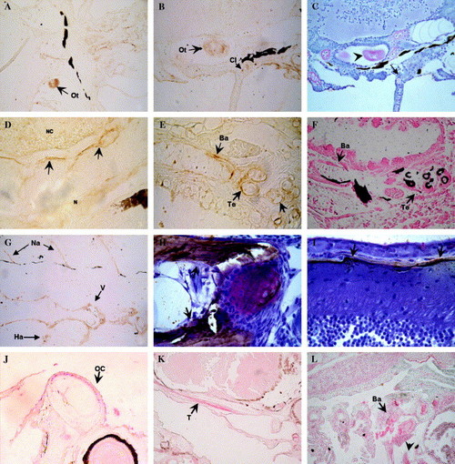
Immunohistochemical detection of Mgp accumulation in zebrafish (A–I) and sole (J–L). (A) Accumulation of Mgp in the otolith at 6 dpf (400×) and (B) in the otolith and cleithrum at 8 dpf (400×); (C) Consecutive section (to that in B) of the otolith (arrowhead) and cleithrum (arrow) stained by Alizarin red and counterstained with toluidine blue (400×); (D) accumulation of Mgp at the zones initiating mineralization of neural arches in pleural vertebrae 3–5 of a 13 dpf larva (1000×); (E) accumulation of Mgp in the mineralizing branchial arches and pharyngeal teeth at 13 dpf; (F) consecutive section (to that in E) stained with von Kossa’s; (G) Mgp accumulation in the forming caudal vertebra and associated arches at 20 dpf (250×); (H) Mgp accumulation in the preopercular bones under endochondral ossification (arrows) in a 40 dpf juvenile (1000×); (I) Mgp accumulation in skull bones undergoing intramembranous ossification (arrow) at 40 dpf (1000×); (J) Mgp accumulation in the otic capsule (OC) at 8 dpf (200×); (K) Mgp accumulation in the mineralizing matrix under the trabecula (T) of a 17 dpf larva (400×); (L) Mgp accumulation in the mineralizing branchial arches and pharyngeal teeth of a 26 dpf (200×), but also in the aorta and cardiac arterial bulbus (arrowhead). No staining was observed in control sections incubated with pre immune serum (results not shown). EXPRESSION / LABELING:
|
|
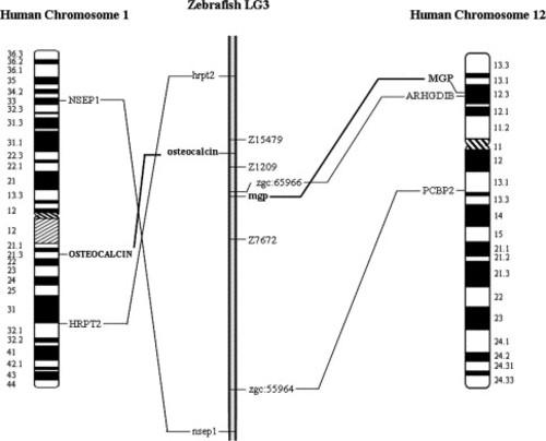
Chromosomal localization of zebrafish mgp and bgp using the LN54 panel. mgp and bgp map on LG3, at 9.54cR from Z1209 and 3.87cR from Z15479 markers, respectively. Some synteny was observed between zebrafish LG3 and human chromosomes 1 and 12, as indicated by lines between syntenic homologues. |
|
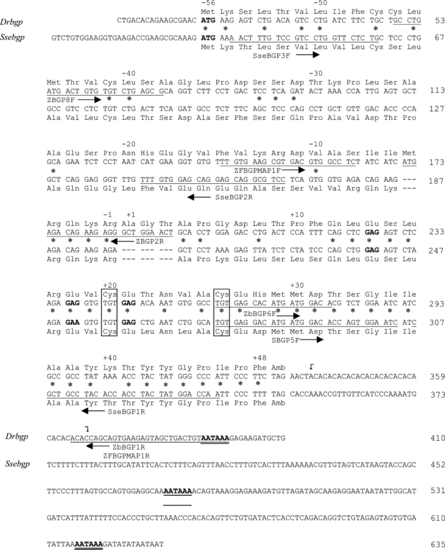
Zebrafish and sole bgp cDNAs. Nucleotide sequence of the cDNAs encoding zebrafish and sole Bgp. The bgp cDNAs were obtained by a combination of RT- and 5′ RACE-PCR amplification. Numbering on right side corresponds to nucleotides. Amino acid residues are numbered according to the first residue of the mature protein and are shown above (for zebrafish) or below (for sole) the corresponding codons in cDNA sequence. The polyadenylation signals are underlined twice and the stop codons are identified by their three letter code. Conserved cysteine residues are boxed. An asterisk is used to show identity between zebrafish and sole amino acid sequences. The sequences used for constructing primers are underlined and marked by horizontal arrows next to the name of the primer. Presumed Gla residues are shown in bold (based in homology with other bgp sequences). A 16 (AC) repetitive motif on zebrafish bgp 3′-UTR region is marked between curved arrows. |
|
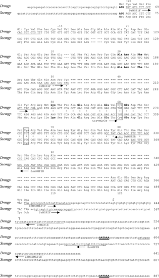
Zebrafish and sole mgp cDNAs. Nucleotide sequence of the two mgp cDNAs were obtained by a combination of RT- and 5′ RACE-PCR amplification. Numbering and labeling is as described in Figure 2s. Amino acid residues are numbered according to the first residue of the mature protein and are shown above the corresponding codon in the DNA sequence, for zebrafish. A 13 (GT) repetitive motif on zebrafish mgp 3′-UTR region is marked between curved arrows. |
|
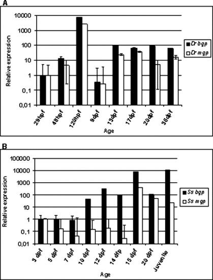
Zebrafish and sole bgp and mgp gene expression detected by real time PCR. Quantitative detection of bgp and mgp mRNA levels throughout development. The relative expression levels were determined with respect to the youngest stage analyzed and are presented as bar graphs using logarithmic scales. (A) zebrafish, (B) sole. EXPRESSION / LABELING:
|
Reprinted from Gene expression patterns : GEP, 6(6), Gavaia, P.J., Simes, D.C., Ortiz-Delgado, J.B., Viegas, C.S., Pinto, J.P., Kelsh, R.N., Sarasquete, M.C., and Cancela, M.L., Osteocalcin and matrix Gla protein in zebrafish (Danio rerio) and Senegal sole (Solea senegalensis): Comparative gene and protein expression during larval development through adulthood, 637-652, Copyright (2006) with permission from Elsevier. Full text @ Gene Expr. Patterns

