- Title
-
Mutations in connexin43 (GJA1) perturb bone growth in zebrafish fins
- Authors
- Iovine, M.K., Higgins, E.P., Hindes, A., Coblitz, B., and Johnson, S.L.
- Source
- Full text @ Dev. Biol.
|
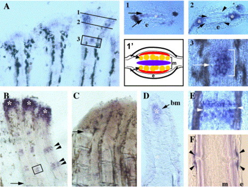
Expression of connexin 43 in fins. In situ hybridization was completed using a probe that recognizes the cx43 coding sequence. (A) Ontogenetically growing fin. Numbers indicate areas of further interest. (1) Transverse section through distal crescent. Arrows, actinotrichia; e, epidermis; *, cx43-expressing cells. (1′) Cartoon of transverse section through the distal crescent. Arrows, actinotrichia; e, epidermis; m, mesenchyme. Blue reflects undifferentiated cells medial to actinotrichia. Red represents cells which have crossed actinotrichia and become determined as osteoblasts (i.e., evx1-positive cells). (2) Transverse section through lateral cx43-stained cells. Arrows, actinotrichia; arrowheads, lepidotrichia; e, epidermis; *, cx43-expressing cells. (3) High magnification of a representative young joint, similar to boxed region in A. Arrow points to newly forming joint. Bracket identifies cx43-stained cells. (B) Wild-type regenerating fin at 5 dpa. Arrow, amputation plane; arrowheads, cx43-stained cells around mature joint; *, cx43-expressing cells in the blastema. (C) sofb123 regenerating fin at 5 dpa. Arrow, amputation plane. (D) Longitudinal cross-section through a cx43-stained regenerating fin. The basement membrane (bm) separates the epithelial and mesenchymal compartments. *, cx43-expression. (E) High magnification of a representative mature joint, similar to boxed region in B. Arrowheads point to joint. (F) Longitudinal cross-section through a mature joint. Arrowheads, cx43-stained cells; e, epidermis; m, mesenchyme. EXPRESSION / LABELING:
|
|
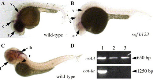
Expression of connexin 43 in embryos. In situ hybridization was completed using a cx43 probe against the cx43 3′ UTR. (A) Expression of cx43 in wild-type embryo at 24 hpf. e, eyes; c, cerebellum; v, vasculature. (B) Expression of cx43 in sof embryo at 24 hpf. e, eyes; c, cerebellum; v, vasculature. (C) Expression of cx43 in wild-type larvae at 72 hpf. Embryos were treated with PTU to prevent the production of melanin and facilitate the identification of stained structures. l, lens epithelium; h, hindbrain; t and arrowhead, thymus. (D) RT-PCR from 72 hpf whole embryos (lane 1), 72 hpf embryonic hearts (lane 2), and adult hearts (lane 3). The top panel shows amplification from cx43-3′ UTR primers, the bottom panel shows amplification from col-1a primers. Approximate sizes of amplified products are shown on the right (arrowhead). |
|
cx43-MO injections cause defects in heart morphology and hematopoiesis. Representative photographs of each morpholino injection trial are shown. (A–C) Wild-type embryos injected with 1.5 ng of standard control morpholino. (D–F) Wild-type embryos injected with 1.5 ng of cx43-MO. (G–I) Wild-type embryos injected with 0.25 ng of cx43-MO. (J–L) sofb123 embryos injected with 0.25 ng cx43-MO. The left column (heart) shows the heart morphology phenotype at 72 hpf. The atrium is outlined in purple, and the ventricle is outlined in green. The middle column (heart) shows higher magnification views of the hearts in the left column. The right column (blood) shows o-dianisidine staining of globin (representing red blood cells) in injected embryos at 72 hpf. |
|
Cell proliferation during ontogenetic growth in wild-type and sofb123 caudal fins. Wild-type and sofb123 fish were labeled with BrdU for 6 h. BrdU-positive cells are labeled in red. The bracket indicates the region from which cells were counted, n indicates the number of BrdU-positive cells in this particular fin ray (note: all BrdU-positive cells are not in this focal plane). Left: wild-type fin ray showing BrdU-labeled cells. On average, 32 ± 5 cells are labeled in the third fin ray from either lobe (n = 10 fins). Right: sofb123 fin ray showing BrdU-labeled cells. On average, 21 ± 3 cells are labeled in the third fin ray from either lobe (n = 10 fins). |
Reprinted from Developmental Biology, 278(1), Iovine, M.K., Higgins, E.P., Hindes, A., Coblitz, B., and Johnson, S.L., Mutations in connexin43 (GJA1) perturb bone growth in zebrafish fins, 208-219, Copyright (2005) with permission from Elsevier. Full text @ Dev. Biol.


