- Title
-
Hedgehog signalling is required for correct anteroposterior patterning of the zebrafish otic vesicle
- Authors
- Hammond, K.L., Loynes, H.E., Folarin, A.A., Smith, J., and Whitfield, T.T.
- Source
- Full text @ Development
|
Expression of Hedgehog pathway components in the zebrafish otic vesicle. (A) Tracing of transverse section, outlining relevant tissues: ot.v, otic vesicle; fp, floorplate; n, notochord; hb, hindbrain. Dorsal towards the top. Scale bar: 50 μm. (B-H) Transverse, hand cut sections of whole-mount in situ hybridisation. Note that midline sources of Hedgehog are approximately 40 μm from the otic vesicle; shh is expressed in both the floorplate and notochord (C), twhh in just the floorplate (E) and ehh in just the notochord (G). Factors necessary for transduction of the Hh signal are expressed within the otic vesicle; ptc1 is expressed in a ventromedial domain (arrow, B), ptc2 throughout ventral otic regions (D) and smo throughout the entire vesicle (F). gli2 is not, however, highly expressed in the developing ear (H). (I,J) Dorsal views of whole-mount otic vesicle preparations. Anterior towards the left, lateral towards the top. Scale bar: 50 μm. At 19 hpf (I), ptc1 is expressed throughout the ventromedial otic vesicle but by 22 hpf (J) is concentrated in posterior regions (brackets). |
|
Expression of ptc1 in two Hh pathway mutants and in embryos in which shh RNA has been overexpressed. Dorsal views of 24 hpf whole-mount otic vesicle preparations showing ptc1 expression. Anterior towards the left, lateral towards the bottom. Scale bar: 50 μm. Note that ptc1 expression is much weaker in contf18b homozygotes (B) than in wild-type embryos (A; a sibling of the contf18b homozygote). ptc1 expression is undetectable in the smub641 otic vesicle (C) and is upregulated throughout the vesicle in embryos in which shh has been overexpressed by injection of 100ng/μl shh RNA (D). |
|
The ears of contf18b and smub641 homozygotes display a loss of posterior structures and a duplication of anterior structures. (A-C) DIC images of live ears, focussed at the level of the anterior otolith. Lateral views; anterior towards the left, dorsal towards the top. Both otoliths in con and smu ears (arrowheads, B,C) are small, lateral and ventral, resembling the anterior otolith (arrowhead, A) of wild-type embryos rather than the larger, medial posterior otolith (out of focus in A). Arrows indicate ventral sensory thickenings (maculae) underlying the otoliths. In the wild type, the anterior (utricular) macula lies under the anterior otolith on the ventral floor of the vesicle (A). In smu, a single ventral macula underlies the two small otoliths (B), while in con, a second ventral macula is found at the posterior of the ear (C). (D-I) Confocal images (projections of z-series) of ears stained with FITC-phalloidin to reveal the actin-rich stereocilia of sensory hair cells. (D-F) Lateral views; anterior towards the left, dorsal to top. (G-I) Dorsal views; anterior towards the left, medial towards the top. (D,G) Wild-type pattern. This is similar between 60 hpf and 4 dpf, but the number of hair cells increases in all patches during this time. Note the rounded anterior macula on the ventral floor of the vesicle (arrow) and the irregularly shaped posterior macula on the medial wall (arrowhead). Asterisks indicate the three cristae. In con and smu, the posterior macula is absent from the medial wall. In smu, a single ventral macula covers the ventral surface of the ear (arrow, E,H). In con, the anterior macula is present as normal (left arrow, F,I), but a second ventral macula is present at the posterior of the ear (right arrow, F,I). This resembles the posterior macula in shape but is smaller than normal. In a proportion of con and smu ears four cristae are present (E). (F,I) con ears with only two cristae, because of the relative immaturity of these ears. (J-L) Transverse paraffin sections (10 μm) through the otic vesicles, stained with Haematoxylin and Eosin. Dorsal towards the top. ot.v, otic vesicle; arrows, ventral maculae; arrowheads, posterior (medial) maculae; asterisks indicate cristae. Midline tissue is lost between the otic vesicles of smu and con embryos, so that the vesicles turn inwards towards the midline. However, all sensory patches are ventral; none are found on the medial wall. Scale bars: 50 μm. |
|
Gene expression in contf18b and smub641 ears. Whole-mount in situ hybridisation; anterior towards the left. (A-C,J-O,Y-Aa) Dorsal views, medial towards the top. All other panels are lateral views, dorsal towards the top. Anterior otic expression domains of otx1 (A-C), wnt4 (D-F) and nkx5.1 (G-I), but not pax5 (J-L) and fgf8 (M-O), are duplicated at the posterior of smu and con otic vesicles. Arrowhead (A,B) shows axis of otx1 symmetry in smu ears. Arrow (A,C) shows axis of otx1 symmetry in con ears. nkx5.1 expression in the statoacoustic ganglion (g) is not duplicated at the posterior of the ear (G-I). Posterior expression domains of follistatin (arrowhead, P-R) are lost. Expression of dorsal (dlx3b, S-U), ventral (eya1, V-X) and medial (pax2a, Y-Aa) markers are not affected in con and smu. An ectopic expression domain of the crista marker msxc is present at the posterior of ∼31% con and 50% smu mutants (Ab-Ad). ot.v, otic vesicle; g, statoacoustic ganglion. Scale bars: 50 μm (shown in the left-hand panel of each set). PHENOTYPE:
|
|
Overexpression of ptc1 RNA in wild-type and contf18b embryos phenocopies the anteriorised ear phenotype seen in contf18b and smub641 homozygotes. Lateral views; anterior towards the left, dorsal towards the top. (A,E,I) DIC images of live embryos, focussed at the level of the anterior otolith. Both otoliths in the ears of ptc1-injected embryos (E,I) are small, lateral and ventral, and resemble the anterior otolith of wild-type embryos (A). (B,F,J) Confocal images of FITC-phalloidin stained ears. (B) Wild-type pattern: arrowhead, anterior macula; arrow, posterior macula. A posterior macula is not present on the medial wall of ptc1-injected embryos (F,J). In ears of ptc1 injected wild-type embryos, the anterior macula is present as normal but a second ventral macula is present at the posterior of the ear, as in con (arrowheads, F). In ears of ptc1-injected embryos from a con/+ mating, a single ventral macula covers the ventral surface of the otic vesicle, as in smu ears (arrowhead, J). (C,D,G,H) In situ markers show similar expression patterns in the ears of ptc1-injected embryos and of con and smu. Four cristae may develop (G), and expression of the anterior marker nkx5.1 is expanded (H). We do not have markers that distinguish between con-like and smu-like ears and so these assays were not repeated on ptc1-injected contf18b embryos. Scale bars: 50 μm. |
|
Anteriorised ear phenotypes in cyc;syu double mutants and twhh antisense morpholino-injected syut4 embryos. Lateral views; anterior towards the left, dorsal towards the top. (A,D,G,J) Wild-type ear pattern; A is taken from Fig. 3 for comparison. (B,E,H,K) Ears of syu;cyc double mutants. (C,F,I,L) Ears of syut4 mutant embryos injected with 0.25mM twhh morpholino. (A-C) DIC images of live ears. The ears of cyc;syu mutants and twhh MO-injected embryos have two small, lateral otoliths (arrowheads B,C) resembling the anterior otolith of ears from wild-type embryos (arrowhead, A). Thickened sensory epithelium is present at both the anterior and the posterior of the vesicle in cyc;syu and syu +twhh MO embryos (arrows, B,C) rather than just at the anterior as in the wild-type (arrow, A). (D-F) Confocal images of FITC-phalloidin stains. Hair cells on the ventral floor are present at both the posterior and anterior of the vesicle in cyc;syu and syu +twhh MO embryos (arrows, E,F) rather than just at the anterior as in the wild-type (arrow, D). The posterior macula (arrowhead, D) is missing from the medial wall in cyc;syu and syu + twhh MO ears (E,F). Four cristae (*) rather than the usual three are present in some cyc;syu and syu + twhh MO embryos (e.g. E). Three cristae were present in the ear shown in F, but only one is in the focal plane. (G-L) In situ hybridisation. Arrows indicate the posterior domain of follistatin expression in the wild type (G). This is absent in cyc;syu and syu+twhh MO ears (H,I). Anterior nkx5.1 expression (J) is expanded in cyc;syu and (less extensively) in syu+twhh MO ears (K,L). Scale bars: 50 μm. PHENOTYPE:
|
|
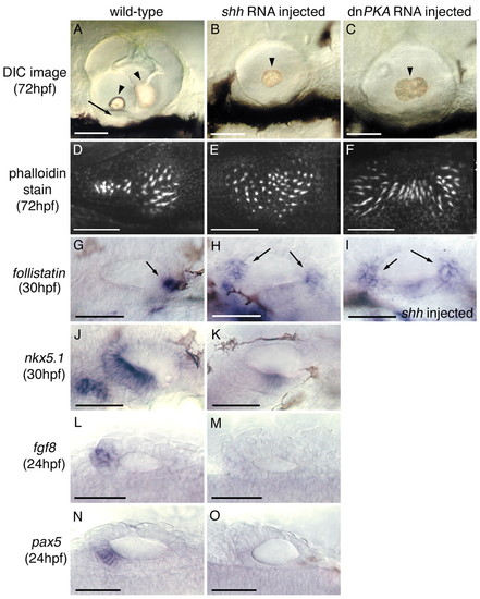
Overexpression of shh and dnPKA RNA in wild-type embryos results in posteriorised ears. (A-K) Lateral views; anterior towards the left, dorsal towards the top. (L-O) Dorsal views; anterior towards the left, lateral towards the top. (A-C) DIC images of live ears. Scale bars: 50 μm. The ears of shh- and dnPKA-injected embryos are small and contain either a single otolith (arrowhead, B) or fused otoliths (arrowhead, C), positioned medially. A ventral anterior macula (arrow, A) is absent in these posteriorised ears. (D-F) Confocal images of FITC-phalloidin stains showing the pattern of sensory hair cell stereocilia in posterior maculae. Scale bars: 25 μm. (D) Wild-type pattern. The posterior macula has a rounded posterior region and a slim anterior projection (D). Posterior maculae of dnPKA-and shh-injected embryos have two rounded posterior ends resulting in a `butterfly′ (E) or `bow-tie′ (F) shape. Note that both phenotypes shown in B,C,E,F could be caused by either shh or dnPKA RNA injection. (G-O) In situ hybridisation performed on shh-injected embryos. Scale bars: 50 μm. (G-I) Posterior follistatin exprcssion is either duplicated at the anterior of the otic vesicle or extends medially along the length of the otic vesicle in posteriorised ears (arrows). (J,K) Anterior nkx5.1 expression is reduced to a small central domain in posteriorised ears. (L-O) Anterior fgf8 and pax5 expression is absent from posteriorised ears. |

Unillustrated author statements PHENOTYPE:
|


