
Image
|
Figure Caption
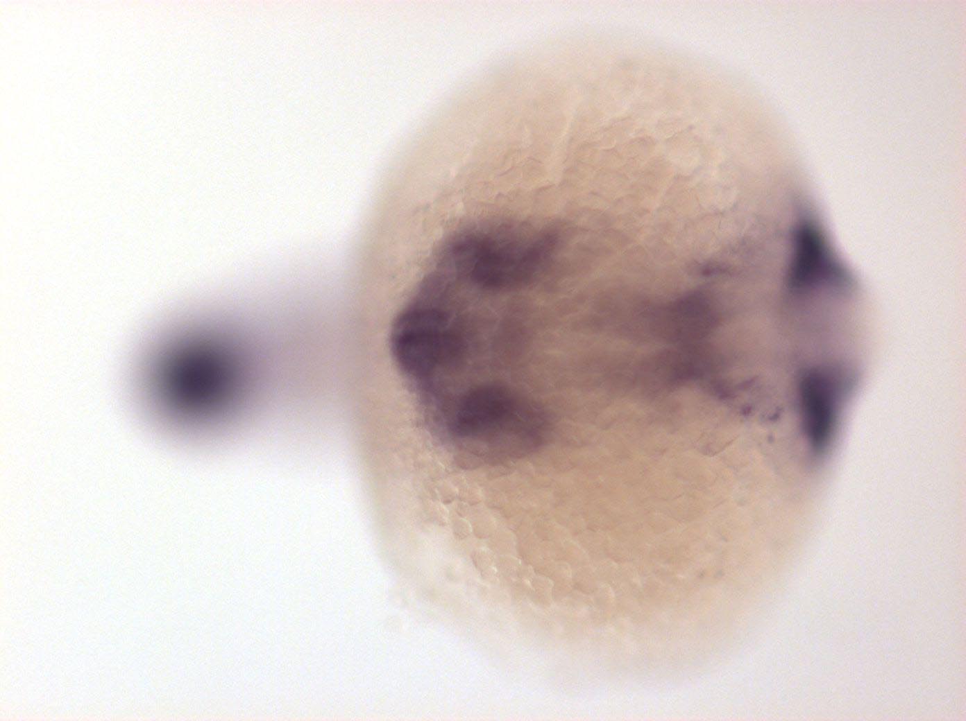
Fig. 3 Expressed in telencephalon, proximal part of retina, midbrain hindbrain boundary, posterior branchial arches, dorsal spinal cord neurons, posterior most spinal cord, pronephric ducts, mucous cells, tail bud and posterior notochord
Developmental Stage
14-19 somites
Orientation
| Preparation | Image Form | View | Direction |
| whole-mount | still | dorsal | anterior to left |
Figure Data