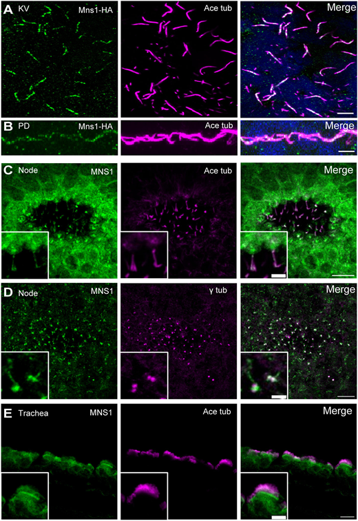
Fig. 2 MNS1 localisation to motile cilia in zebrafish and mouse tissues. (A) Immunofluorescence with antibodies to HA epitope (green) and acetylated tubulin (magenta) of cilia in zebrafish KV [10-somite stage (14 hpf)]. (B) Immunofluorescence with antibodies to HA (green) and acetylated tubulin (magenta) of zebrafish pronephric duct (PD) cilia (24 hpf). (C) Immunofluorescence with antibodies to MNS1 (green) and acetylated tubulin (magenta) of E8.0 mouse node cilia. (D) Immunofluorescence with antibodies to MNS1 (green) and γ tubulin (magenta) of E8.0 mouse node cilia. (E) Immunofluorescence with antibodies to MNS1 (green) and acetylated tubulin (magenta) of adult mouse trachea. Insets in C-E show higher magnification views. Scale bars: 5 µm (A,B,E, main panel); 2 µm (C,D, main panels); 10 µm (C-E, insets).
Image
Figure Caption
Acknowledgments
This image is the copyrighted work of the attributed author or publisher, and
ZFIN has permission only to display this image to its users.
Additional permissions should be obtained from the applicable author or publisher of the image.
Full text @ Development

