IMAGE

Fig. 1

ID
ZDB-IMAGE-240624-29
Source
Figures for İş et al., 2024
Image
Figure Caption

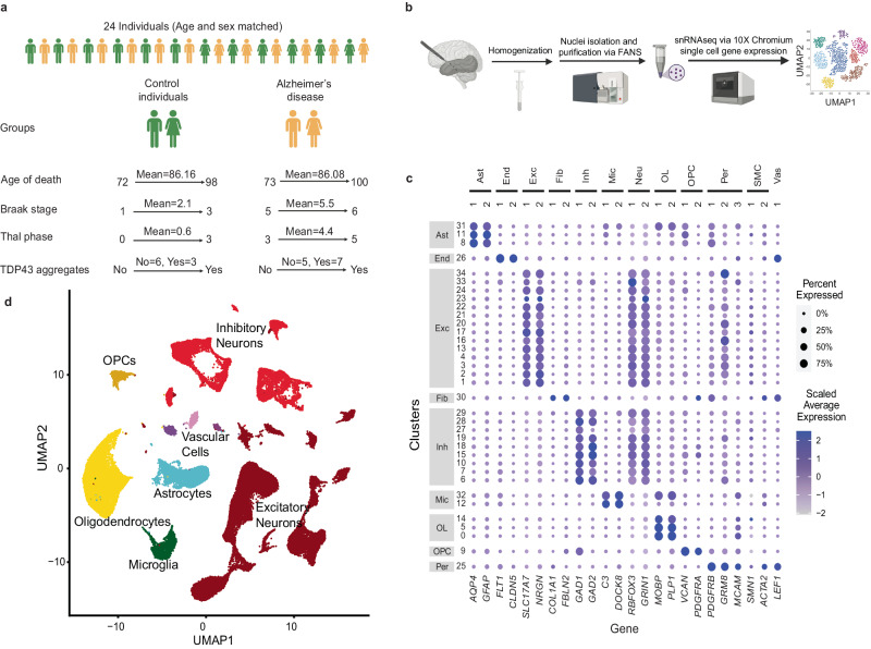
Fig. 1 Summary of the snRNAseq approach utilized in this study.

a Post-mortem temporal cortex tissue from 24 individuals that comprise sex and age matched AD and control individuals were used in this study. b Development and optimization of nuclei isolation protocol for snRNAseq platform. c Well-established cell type markers were used to annotate nuclei clusters and d major brain cell types were visualized in UMAP plots. Figure 1/panels a and b Created with BioRender.com released under a Creative Commons Attribution-NonCommercial-NoDerivs license.

Acknowledgments
This image is the copyrighted work of the attributed author or publisher, and ZFIN has permission only to display this image to its users. Additional permissions should be obtained from the applicable author or publisher of the image. Full text @ Nat. Commun.