
|
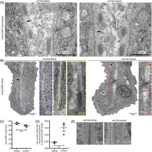
Fig. 10 Actinotrichia of the MFF of wnt10a mutants do not extend properly into the cleft of epidermal cleft cells. All images show TEM micrographs of transverse sections through the ventral MFFs of wnt10a siblings and mutants of 48 hpf, proximately 750 μm anterior of the tip of the caudal fin. (A) In intermediate proximodistal positions, the ventral MFF mutants display regular ultrastructure, including the basement membrane between epidermal cells and the dermal space (arrowheads), basement membrane–dermal anchorage and actinotrichia. (B) In distal-most positions of the ventral MFF, cleft cells of wnt10a mutants display a slightly widened cleft (indicated by blue and red brackets for sibling and mutant, respectively). In addition, in contrast to siblings, in mutants, actinotrichia do not reach the distal tip of the cleft (distal end of actinotrichia in sibling and mutant indicated by blue and red arrowheads, respectively). Furthermore, siblings show actinotrichia extending into the interior of cleft cells, constituting “fibripositor”-like structures71 (indicated by yellow arrow in sibling), whereas mutants do not. Blue, yellow, and red dashed rectangles of overview images frame regions shown in magnified views to their right. (C, D) Graphical illustration of quantification demonstrating that wnt10a mutant embryos do not have significantly shorter ventral MFFs compared to the siblings (C), but a statistically significant increase in the distance between actinotrichia and the distal tip of the cleft of cleft cells (D). The blue dots and red squares in (C, D) indicate values for the respective sibling and mutant shown in (B). (E) At the level of ridge cells, actinotrichia of siblings and mutants display comparable width and banded structure. MFF, median fin fold.