
|
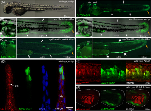
Fig. 6 Canonical Wnt signals are received by distal-most epidermal cells and fin mesenchymal cells of the embryonic MFF. Fluorescent images of live Tg(6xTCF:eGFP) transgenics, with cells receiving canonical Wnt signals in green. (A) At 48 hpf, the responder transgene is expressed in distal-most cells of the MFF (white arrows) and in fin mesenchymal cells (orange arrow) of the caudal fin fold of wild-type embryo. (B) Compared to wild-type sibling (left panel), wnt10a mutant embryo of 60 hpf (right panel) shows reduced Tg(6xTCF:eGFP) expression in the minor and major lobes of the MFF (indicated by arrowheads), whereas expression in the caudal fin fold (indicated by orange arrow) and in the AER of the pectoral fin is comparably unaltered. (C) Compared to non-heat-shocked control (left panel), transgenic embryo with heat-shock-induced global wnt10a overexpression from 24 to 48 hpf (right panel) shows increased Tg(6xTCF:eGFP) expression in the minor and major lobes of the MFF (indicated by thick arrows), whereas expression in the caudal fin fold (indicated by orange arrow) and the AER of the pectoral fin is unaltered. (D) Transverse cryosection approximately 0.5 mm anterior of the caudal tip of the tail of 6xTCF:eGFP 48 hpf embryo, stained with antibodies against GFP and collagen II (labeling actinotrichia, red). The Wnt responder is expressed in distal-most positions of the MFF. (E) 6xTCF:eGFP, GBT0245 (msxc; marker of fin mesenchymal cells in red55) double transgenic embryo revealing expression of the Wnt responder in fin mesenchymal cells of the ventral MFF at 3 dpf. (F) At the onset of metamorphosis (13 dpf, SL 5 mm), the Wnt responder is expressed distal of forming lepidotrichia (stained with alizarin red) of the caudal fin. act, actinotrichia; AER, apical ectodermal ridge; ds, dermal space; MFF, median fin fold; otv, otic vesicle; pecf, pectoral fin; SL, standard length.