
|
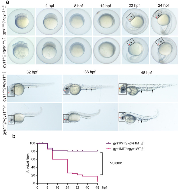
Fig. 1 Maternal gys1 mutant embryos exhibit morphological defect and high mortality rate. (a) Bright-field images showing delayed embryonic development of maternal gys1 mutant embryos from gys1−/− ♀ cross with gys1+/+ ♂, in contrast to time-matched parental gys1 mutant embryos from gys1−/− ♂ cross with gys1+/+♀ from 2 hpf to 48 hpf. Small head phenotype was indicated in rectangles, brain necrosis was indicated with red arrows, small eye phenotype was indicated with asterisks, reduced pigmentation phenotype was indicated with black arrows. (b) Survival Rate of embryos from gys1−/− ♀ cross with gys1+/+ ♂ (n = 1175) and gys1−/− ♂ cross with gys1+/+♀ (n = 885) from 0 hpf to 48 hpf, survival rate was plotted by Kaplan-Meier method, Log-rank (Mantel-Cox) test showed p < 0.0001. (For interpretation of the references to colour in this figure legend, the reader is referred to the Web version of this article.)