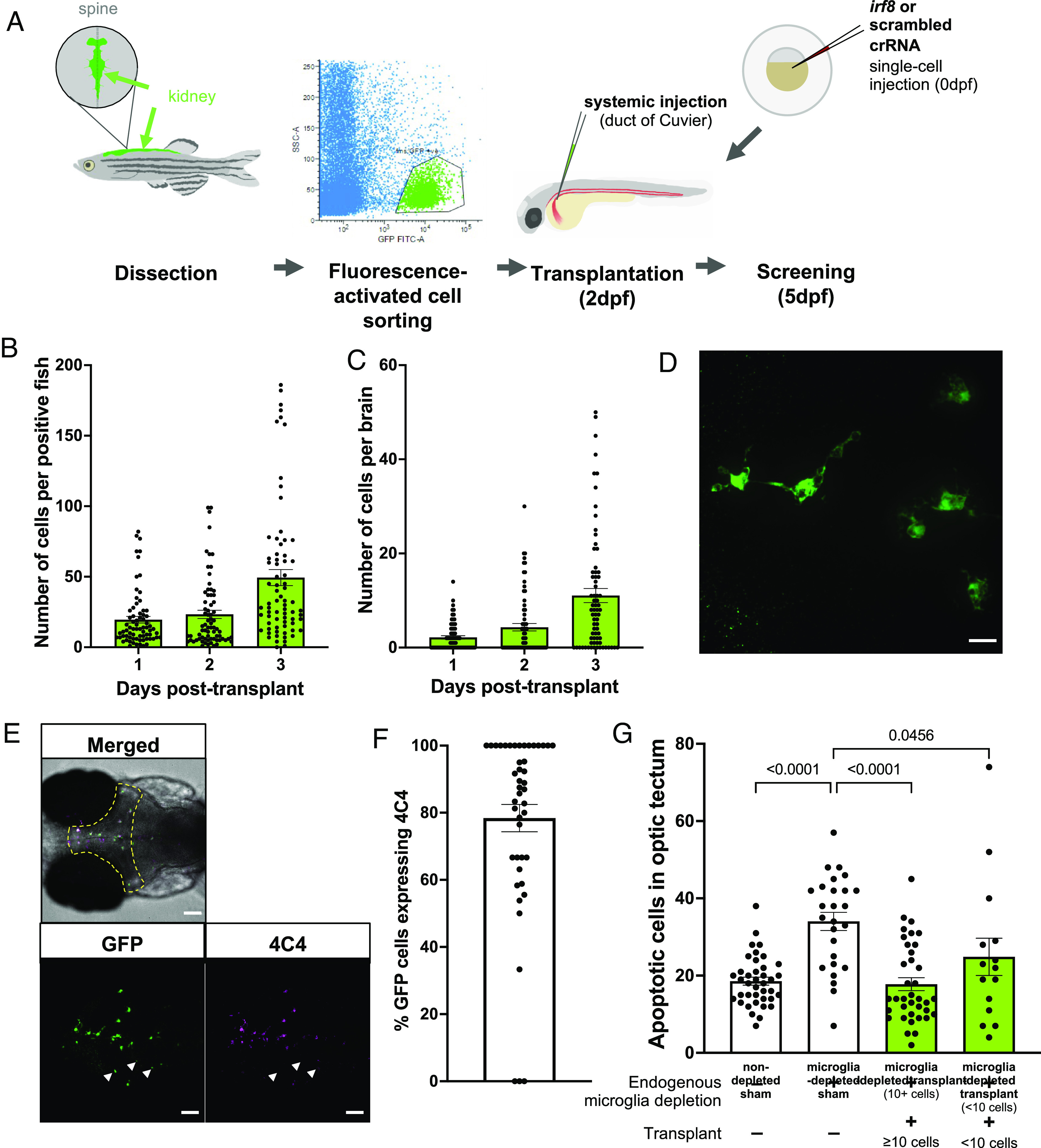
Macrophage transplantation successfully replaces microglia in WT zebrafish. (A) Transplants were performed by dissecting kidneys from Tg(fms:GFP) adult donors and isolating GFP-positive cells using fluorescence-activated cell sorting. Cells were injected into 2 dpf Tg(mpeg1:mCherryCAAX)sh378 hosts via duct of Cuvier. Hosts had previously undergone microglia depletion via targeting of irf8 by CRISPR/Cas9 or were nondepleted with a scrambled control. (B and C) The number of fms:GFP cells per successfully transplanted animal increased throughout the whole body (B) and within the brain (C) across 3 d post-transplant, as revealed by manual counting. (D) Confocal imaging reveals that transplant-derived cells show a branched, microglia-like morphology. The scale bar represents 17 µm. (E and F) Transplant-derived cells express the microglia-specific marker 4C4 at 3 d post-transplant (5 dpf). Colocalization was assessed by manual counting. Arrows indicate GFP-positive cells that do not co-localize with 4C4. The yellow dashed line indicates the border of the optic tectum (region of interest). Three biological replicates, n = 45. The scale bar represents 75 µm. (G) TUNEL staining reveals that transplantation can rescue the number of uncleared apoptotic cells in microglia-depleted brains in a dose-dependent manner, as revealed by manual counting of cells within optic tectum as in E. Kruskal–Wallis test with Dunn’s multiple comparisons. Four biological replicates, n = 15 to 39.
Acknowledgments
This image is the copyrighted work of the attributed author or publisher, and
ZFIN has permission only to display this image to its users.
Additional permissions should be obtained from the applicable author or publisher of the image.
Full text @ Proc. Natl. Acad. Sci. USA
Your Input Welcome
Thank you for submitting comments. Your input has been emailed to ZFIN curators who may contact you if
additional information is required.
Oops. Something went wrong. Please try again later.