IMAGE

Fig 4

ID
ZDB-IMAGE-240511-4
Source
Figures for Ahuja et al., 2024
Image
Figure Caption

Fig 4 cxcl12b is regulated by nkx3.1; loss of Cxcr4 signaling reduces brain pericytes.

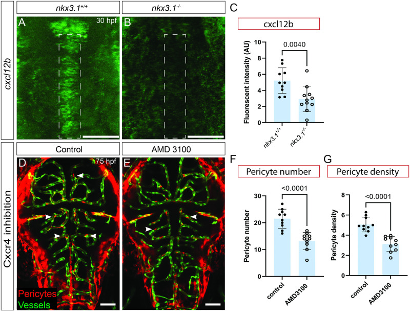
All embryos were imaged dorsally in the head region. cxcl12b mRNA expression is reduced in the embryonic head of nkx3.1−/− mutants (B) as compared to wild-type controls (A) at 30 hpf as quantified by fluorescent intensity (C) of regions marked by dotted lines in A and B. (n = 10 wild-type and 10 mutant embryos). Inhibition of Cxcr4 from 24–75 hpf using AMD 3100 leads to reduced brain pericytes in treated (E) vs. untreated (D) embryos (at 75 hpf (n = 10)). Pericytes (red cells, white arrows) are labeled by TgBAC(pdgfrβ:Gal4); Tg(UAS:NTR-mCherry). Brain vessels in D and E are labeled by Tg(flk:GFP; green). Quantification of (F) pericyte number and (G) pericyte density. (n = 10 wild type and 10 mutants). Statistical significance was calculated using the Student t test. Scale bar is 50 μm. The data underlying this figure can be found in S3 Table.

Acknowledgments
This image is the copyrighted work of the attributed author or publisher, and ZFIN has permission only to display this image to its users. Additional permissions should be obtained from the applicable author or publisher of the image. Full text @ PLoS Biol.