IMAGE

Fig. 1

ID
ZDB-IMAGE-240426-85
Source
Figures for Raote et al., 2024
Image
Figure Caption

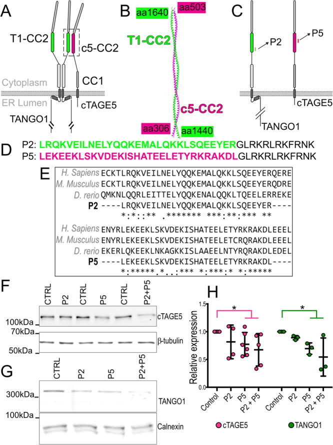
Fig. 1 Design of peptides to inhibit TANGO1-cTAGE5 heterodimerization.

A The organization and the domains of TANGO1 and cTAGE5. Each of these proteins contains two cytoplasmic coiled-coil domains. TANGO1 coiled-coil 2 (T1-CC2) is highlighted in green. cTAGE5 CC2 in magenta. B Alphafold2 prediction of the structure of the heterodimer of TANGO1 (green) and cTAGE5 (magenta). C Schematic indicating where the peptide sequences map to in TANGO1 and cTAGE5. D Two peptides were synthesized of 30 (P2) and 31 (P5) amino acids respectively, conjugated to a 12 amino-acid C-terminal lysosomal escape motif (shown in black text). E Alignment of human, mouse, and zebrafish sequences. F Representative western blot of U2OS cell lysates treated with P2, P5 or P2 + P5, probed for cTAGE5. β-tubulin was used as a loading control. G Representative Western blot of U2OS cell lysates treated with P2, P5 or P2 + P5, probed for TANGO1. Calnexin was used as a loading control. H Quantification of blots showing TANGO1 (green) and cTAGE5 (magenta) levels (mean +/- SD) in control or peptide-treated cells (normalized to control) *p < 0.05, Student’s t test comparing cTAGE5 (magenta line) or TANGO1 (green line) in control vs treated. N = 3 (TANGO1), N = 5 (cTAGE5).

Acknowledgments
This image is the copyrighted work of the attributed author or publisher, and ZFIN has permission only to display this image to its users. Additional permissions should be obtained from the applicable author or publisher of the image. Full text @ Nat. Commun.