
|
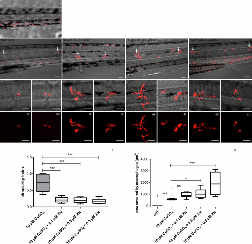
Fig. 4 A set of images and a graph documenting changes in the phenotype of macrophages recruited to 10 μM CuSO4 derived damaged hair cells of L2, L3 or L4 neuromast resulting from co-treatment of 3 dpf Tg(mpeg1:mcherry) larvae with XN. a) The control larva presented normal distribution of macrophages which were found in the ventral myotomes of the trunk and tail. b) 40 min 10 μM CuSO4 exposure triggered macrophages recruitment towards damaged hair cells, as well resulted in a round shape of immune cells. (c,d, e) XN co-treatment did not altered macrophage recruitment, but resulted in the phenotype switch to the ameboid one. White arrows indicate macrophages clustered around injured L2, L3 or L4 neuromasts. Images with 1 and 2 numbers correspond to the main images and show single neuromasts under 2× magnification. Images with prims show the same elements on the black background for better visualization. The visualization was accomplished using a Zeiss LSM-700 confocal microscope (10× objective). (f) the graph presenting the influence of XN on the total area covered by the macrophages associated with L2, L3 or L4 neuromasts. (g) the graph presenting the influence of XN on CI of macrophages recruited to L2, L3 or L4 neuromasts (one-way ANOVA, GraphPad Prism 5, *** p < 0.001, ** p < 0.01, * p < 0.05, ns –statistically not significant). Scale bar = 50 μm (a-e), scale bar = 25 μm (images with 1 and 2, and prims).