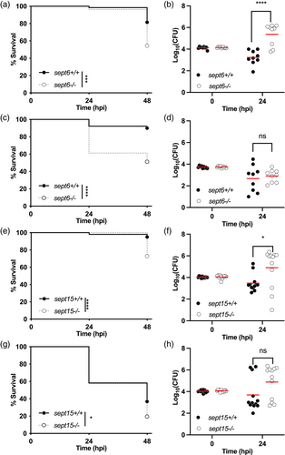
Fig. 3 Zebrafish null mutants of Sept6 and Sept15 are more susceptible to Shigella infection. (a–d) Survival curves (a,c) and Log10-transformed CFU counts (b,d) of 3 dpf sept6+/+ and sept6−/− larvae (wild-type in black; mutant in white) injected in the hindbrain ventricle (HBV) with S. flexneri GFP+ at a dose of 10,000 CFUs (a,b) or a dose of 5,000 CFUs (c,d). Next, larvae were incubated at either 28.5°C (a,b) or 32.5°C (c,d), for up to 48 h. Experiments are cumulative of three biological replicates. Sample size: For survival analysis, a total of 65 (wild-type) and 79 (mutant) and 90 (wild-type) and 80 (mutant) larvae was analysed at 28.5°C and 32.5°C, respectively. For CFU analysis, a total of 9 larvae was analysed per experimental group. Only living larvae were used for CFU enumeration. Statistics: Log-rank (Mantel-Cox) test (a,c); unpaired t test on Log10-transformed values (b,d). ****p < 0.0001; ns p > 0.05, respectively. (e,h) Survival curves (e,g) and Log10-transformed CFU counts (f,h) of 3 dpf sept15+/+ and sept15−/− larvae (wild-type in black; mutant in white) injected in the HBV with S. flexneri GFP+ at a dose of 10,000 CFUs. Next, larvae were incubated at either 28.5°C (e,f) or 32.5°C (g,h) for up to 48 h. Experiments are cumulative of four biological replicates. Sample size: For survival analysis, a total of 99 (wild-type) and 104 (mutant) and 103 (wild-type) and 113 (mutant) larvae was analysed at 28.5°C and 32.5°C, respectively. For CFU analysis, a total of 12 larvae was analysed per experimental group. Only living larvae were used for CFU enumeration. Statistics: Log-rank (Mantel–Cox) test (e, g); unpaired t test on Log10-transformed values (f,h). *p < 0.05; ****p < 0.0001; ns p > 0.05, respectively
Image
Figure Caption
Figure Data
Acknowledgments
This image is the copyrighted work of the attributed author or publisher, and
ZFIN has permission only to display this image to its users.
Additional permissions should be obtained from the applicable author or publisher of the image.
Full text @ Cytoskeleton

