IMAGE

Figure 1

ID
ZDB-IMAGE-231204-35
Antibodies
Source
Figures for Serrano et al., 2023
Image
Figure Caption

Figure 1

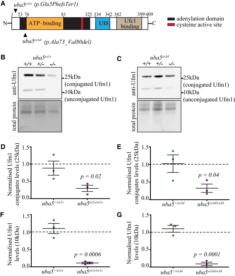
Ufm1 and Ufm1-conjugate levels are reduced in uba5−/− fish at 6 dpf. (A) Schematic of Uba5 wild-type protein and mutation sites in zebrafish. The mutant strains were generated using CRISPR/Cas9 genome editing. The uba5ex1s strain carries a 73-base pair insertion following the ATG, resulting in a frameshift and incorporation of a premature stop after 5 amino acids, whereas the uba5ex3d strain carries a 24-base pair deletion that removes the ATP-binding domain. (B–G) Western blot for Ufm1 and Direct Blue staining for total protein (loading control) on protein lysates obtained at 6 dpf. Ufm1 and Ufm1-conjugate levels were normalized to that of the respective total protein levels. The changes in Ufm1 and Ufm1-conjugate levels in uba5 mutants were determined relative to the levels of wild-type siblings. Statistical significance was calculated using a one-way ANOVA with Dunnett’s post hoc multiple comparison correction test. Data are presented as mean ± SEM for three independent biological replicates, each consisting of a pooled sample of 20–25 embryos. UIS, Ufm1-interacting sequence; Ufc1, ubiquitin-fold modifier conjugating enzyme 1; Ufm1, ubiquitin-fold modifier 1.

Figure Data
Acknowledgments
This image is the copyrighted work of the attributed author or publisher, and ZFIN has permission only to display this image to its users. Additional permissions should be obtained from the applicable author or publisher of the image. Full text @ Brain Commun