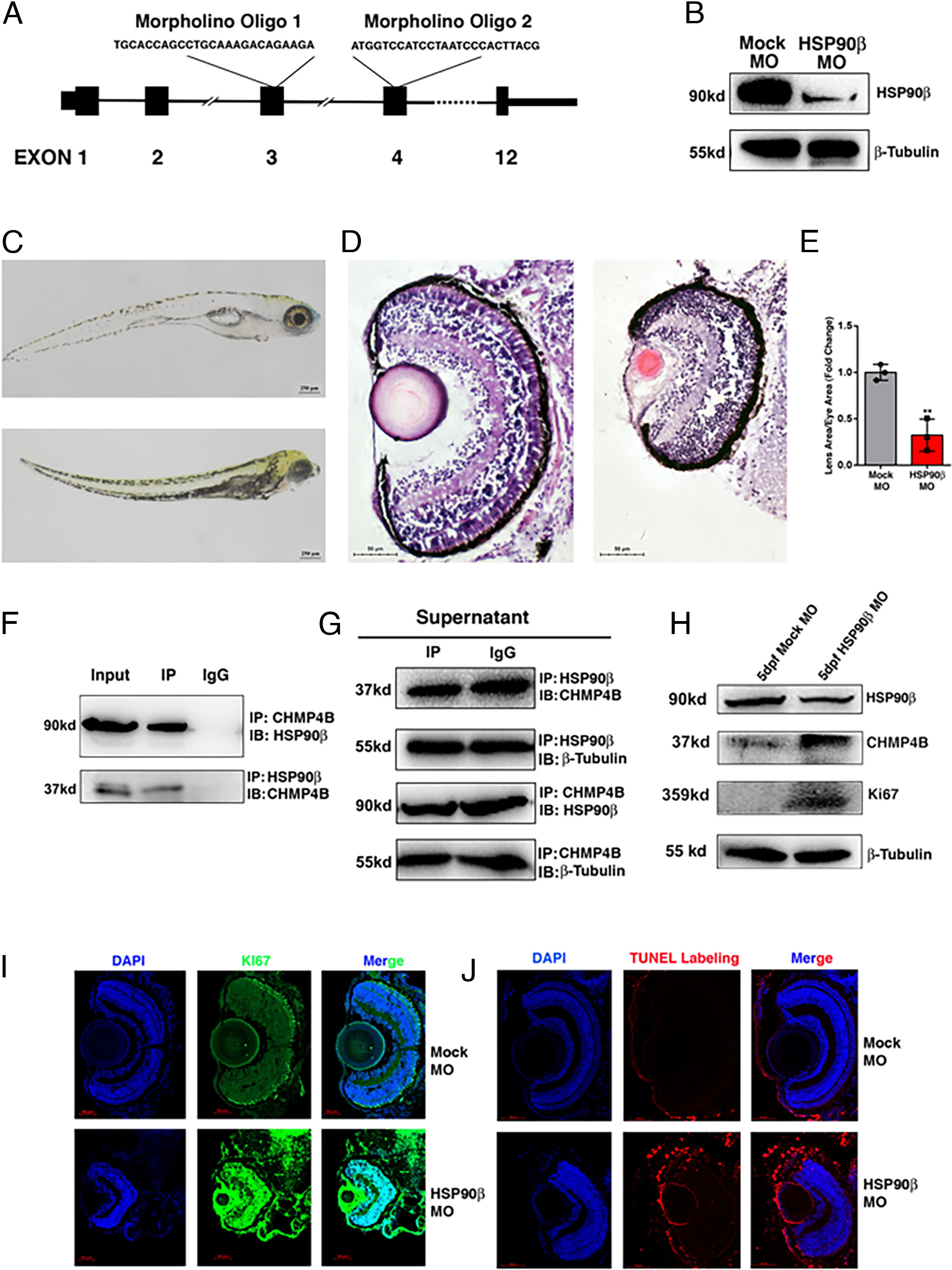
Fig. 3 Silence of HSP90βcauses cataractogenesis in zebrafish lens. (A) Two morpholino oligos targeting exon 3 and exon 4 in Zebrafish were designed to silence HSP90β expression. (B) Western blot analysis of HSP90β levels in mock-MO and HSP90β-MO treated 5-d postfertilization (dpf) zebrafish embryo. (C) Morphology of 5 dpf mock-MO (Top) and HSP90β-MO (Bottom) zebrafish larvae. (D) H.E. staining of 5dpf mock-MO (Left) and HSP90β-MO (Right) zebrafish eye by frozen sectioning. (E) Quantification of the ratio between lens and eye size in mock-MO and HSP90β-MO 5dpf zebrafish embryos. (F) Co-IP between HSP90β and CHMP4B, endogenous HSP90β and CHMP4B were precipitated by CHMP4B and HSP90β antibodies, respectively, and detected by western blot analysis. Input protein [0.05% (Top) and 5% (Bottom)] were included as control. (G) For comparison, supernatant samples of IP were also analyzed by western blot. (H) Western blot analysis showed that the expression levels of CHMP4B and Ki67 were clearly up-regulated when HSP90β was silenced by morpholino oligos in zebrafish. (I) IF staining of Ki67 showed that cell proliferation is up-regulated in 5dpf HSP90β-MO zebrafish eye than in mock-MO zebrafish eye. (J) TUNEL labeling assay was used to detect cell apoptosis in zebrafish eye. Compared to 5dpf mock-MO zebrafish, 5dpf HSP90β-MO zebrafish showed significantly more cell apoptosis in lens epithelium. Error bar represents SD. **P < 0.01.
Image
Figure Caption
Figure Data
Acknowledgments
This image is the copyrighted work of the attributed author or publisher, and
ZFIN has permission only to display this image to its users.
Additional permissions should be obtained from the applicable author or publisher of the image.
Full text @ Proc. Natl. Acad. Sci. USA

