
|
Figure 6
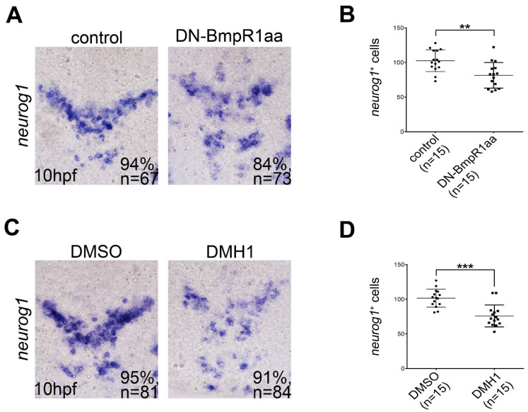
Inhibiting BMP signaling reduces the formation of neuronal precursors. All panels show dorsal views of flat-mounted embryos with the anterior on the top. (A,C) In situ hybridization of 10 hpf embryos focusing on the midbrain and hindbrain regions showing that neurog1-positive cells were reduced by heat-shock-induced DN-BmpR1aa (A) or DMH1 treatment (C) at 8 to 10 hpf. This was quantified by the cell-count analysis shown in (B,D), respectively. n, total number of embryos analyzed from three independent experiments. The percentages in each panel in (A,C) indicate the proportion of embryos displaying the same phenotype as that shown in the photographs of the total embryos examined. Quantitative data are presented as mean ± standard deviation (SD). ** p < 0.01; *** p < 0.001.