Fig. 3
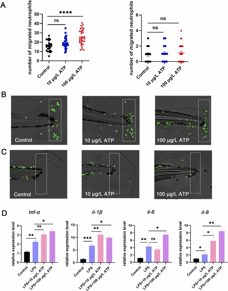
Fig. 3. Inhibition of mAChRs up-regulated neutrophils migration and increased proinflammatory cytokines level. (A) Neutrophils were photographed with a fluorescence microscope (n = 25), and neutrophils migration increased after atropine (0.003 μM, 0.03 μM) treatment in the statistical area. The caudal fin was not injured in the right picture, and the caudal fin was injured in the left picture. (B) Model of caudal fin injury: atropine (0.003 μM, 0.03 μM) treated or untreated caudal fins of zebrafish were cut with a scalpel blade (white rectangles indicate statistical areas). (C) When the zebrafish caudal fin was not damaged, there was no difference in the number of neutrophils in the statistical area between atropine (0.003 μM, 0.03 μM) and the control group (the white rectangles indicate the statistical area). (D) The expression levels of tnf-α, il-1β, il-6 and il-8 in zebrafish treated with atropine (0.003 μM, 0.03 μM) were increased compared with the LPS group (n = 30). (*P < 0.05,** P < 0.01, ***P < 0.001, ****P < 0.0001, one-way ANOVA analysis, LSD for port-hoc test).

