
Image
|
Figure Caption
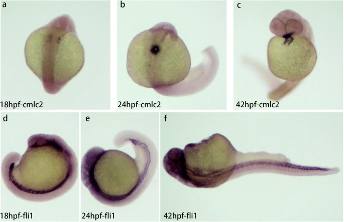
Fig. 1
Expression of cardiomyocyte and endothelial marker genes (cmlc2, fli1) at three different stages of zebrafish embryo development (18, 24, 42 hpf). a-c Expression of cmlc2 at 18hpf (a), 24hpf (b), and 42hpf (c). A pair of primordia were generated at 18 hpf, one on each side of the midline (a), The primordia on both sides fused to form a single linear heart tube at 24 hpf (b), the heart tube undergoes a loop and the heart chamber was visible (c). V, ventricle; A, atrial. d-f Expression of fli1 at 18 hpf (d), 24 hpf (e). and 42hpf (f)
Acknowledgments
This image is the copyrighted work of the attributed author or publisher, and
ZFIN has permission only to display this image to its users.
Additional permissions should be obtained from the applicable author or publisher of the image.
Full text @ BMC Genomics