Image
Figure Caption
Fig. 2
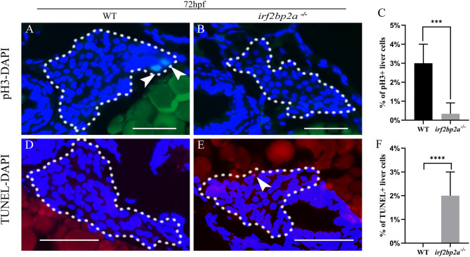
Fig. 2. Deficiency of irf2bp2a leads to decreased proliferation and enhanced apoptosis of liver cells. (A, B, D, E) The proliferation and apoptosis status of hepatic cells were detected by pH3 staining and TUNEL assay in 72 hpf embryos. The sections were counterstained with DAPI to label the nucleus. White dashed lines indicate the liver boundary. White arrows indicate pH3 or TUNEL positive cells. Scale bar: 100 μm. (C, F) Statistical analysis of hepatocyte proliferation and apoptosis. p values are denoted by asterisks. (Student t-test, N ≥ 3. Error bars represent mean ± SD, ***P < 0.001, ****P < 0.0001).
Figure Data
Acknowledgments
This image is the copyrighted work of the attributed author or publisher, and
ZFIN has permission only to display this image to its users.
Additional permissions should be obtained from the applicable author or publisher of the image.
Reprinted from Biochimica et biophysica acta. General subjects, 1866(10), Yan, L., Gao, S., Zhu, J., Zhou, J., Irf2bp2a regulates liver development via stabilizing P53 protein in zebrafish, 130186, Copyright (2022) with permission from Elsevier. Full text @ BBA General Subjects

