IMAGE

Fig. 6

ID
ZDB-IMAGE-220809-6
Source
Figures for Hu et al., 2022
Image
Figure Caption

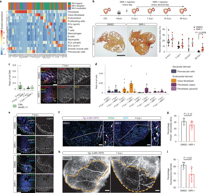
Fig. 6 Cellular dissection of the role of canonical Wnt signalling.

a, Expression of Wnt signalling factors in different cell types of the zebrafish regenerating heart. b, Upper panel: cartoon summary of IWR-1 Wnt inhibition experiments. Lower left: histological comparison of the IA at 30 d.p.i. with intraperitoneal (IP) injections of IWR-1 or DMSO. In the paraffin AFOG-stained sections, fibrin-red and collagen-blue are clearly visible in the IA in IWR-1-treated samples at 30 d.p.i. Scale bar, 300 μm. Lower right: relative size of the IA area as a percentage across all histological replicates; data shown as mean and s.d. Two-tailed unpaired Student’s t test between groups at each time point, P = 0.0059. c, Left: changes in relative numbers of dedifferentiated CMs at 3 and 7 d.p.i. between IWR-1- and DMSO-treated hearts (error bars indicate s.e.m.). Right: localization of dedifferentiated (ttn.2) cardiomyocytes at 7 d.p.i. with and without Wnt inhibition. Scale bar, 100 μm. d, Changes in relative numbers of non-CMs following Wnt inhibition at 3 and 7 d.p.i. (error bars indicate s.e.m.). e, Fluorescence in situ hybridization of perivascular cells (pdgfrb, white) and nppc fibroblasts (nppc, purple) at 7 d.p.i. with and without Wnt inhibition. Scale bar in c and e, 100 μm. White dashed lines indicate the IA. f, Immunostaining of Tg(-0.8flt1:RFP) hearts at 4 d.p.i. with and without Wnt inhibition; RFP (coronaries, magenta), PCNA (proliferation marker, green) and DNA (DAPI, blue). Arrowheads point to PCNA+ cECs, white dashed lines indicate the IA. Scale bar, 100 μm. g, Percentages of PCNA+ cECs in DMSO-injected (n = 5) and IWR-1-injected (n = 6) fish at 4 d.p.i. Data are shown as mean and s.d. Two-tailed unpaired Student’s t test, P = 0.12. h, Whole-mount images of Tg(-0.8flt1:RFP) hearts at 7 d.p.i. with and without Wnt inhibition. Orange dashed lines indicate injury area. Scale bar, 100 μm. i, Percentages of RFP fluorescence intensity in injured tissue of DMSO-injected (n = 5) and IWR-1-injected (n = 5) fish at 7 d.p.i. Data are shown as mean and s.d. Two-tailed unpaired Student’s t test, P = 0.02.

Source data

Acknowledgments
This image is the copyrighted work of the attributed author or publisher, and ZFIN has permission only to display this image to its users. Additional permissions should be obtained from the applicable author or publisher of the image. Full text @ Nat. Genet.