
|
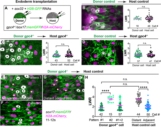
Fig. 2 Gpc4 is required in a cell-autonomous and non-cell-autonomous manner for planar polarity of endodermal cells. (A) Schematic illustrating endoderm transplantation in embryos obtained from incrossing gpc4+/−/sox17:memGFP/H2A-mCherry zebrafish. (B-G) XY view confocal images (Z-projection) showing endoderm in the indicated host embryos transplanted with the indicated donor endoderm cells (the nuclei of which are white, indicated by asterisks). Graphs show average LWR of host (green) and donor (black) endodermal cells at the 12s stage of control donor cells transplanted into sibling control hosts (B, four embryos); gpc4−/− donor cells transplanted into gpc4−/− hosts (C, five embryos); and control donor cells transplanted into gpc4−/− hosts (D, three embryos). (E-G) XY view confocal images (Z-projection) showing endodermal cells displaying different patterns in control hosts transplanted with gpc4−/− cells (nine embryos). Yellow dots show control host cells. (H) Average LWR of host and donor endodermal cells at the 12s stage. All cells analyzed are plotted and the number of cells is shown. Dark green dots, pattern 1 donor cells; light green dots, pattern 2 donor cells; grey dots, distant host cells; blue dots, host cells adjacent to donor cells. Data are mean±s.e.m. n.s., not significant, P>0.05; ****P<0.0001 (unpaired, two-tailed Student's t-test).