
|
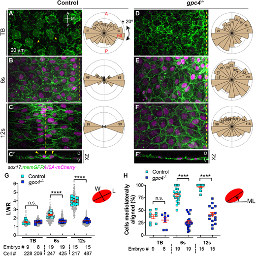
Fig. 1 Gpc4 is required for endodermal cell polarity during segmentation. (A-F′) Confocal images showing endodermal cells with plasma membrane (GFP) and nuclei (pseudo-colored magenta) labeled in the indicated embryos at the tailbud (TB), 6s and 12s stages. (A-F) Z-projection of XY view; (C′,F′) Z-projection of XZ view. Asterisks indicate gaps between cells; dashed-yellow lines indicate the midline. A, anterior; D, dorsal; ML, mediolateral; P, posterior; V, ventral. Rose plots illustrate the cell orientation in indicated embryos (each bin, 20°). (G) Average length-to-width ratio (LWR) of endodermal cells in embryos in A-F. Data from all embryos (squares) and all cells (gray circles) are superimposed, with the number of cells and embryos indicated. (H) Percentage of cells the longitudinal axis of which was oriented ±20° with respect to the ML embryonic axis in embryos in A-F. Data are mean±s.e.m. n.s., not significant, P>0.05, ****P<0.0001 (unpaired, two-tailed Student's t-test).