
|
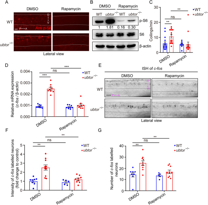
Fig. 5
Rapamycin normalizes spontaneous movements and the activity of spinal interneurons in 28-hpf ubtor mutant embryos. A p-S6 staining in the spinal cord of 28-hpf embryos treated with DMSO (solvent) or 100 nmol/L rapamycin (dashed lines, spinal somites 4–10; scale bar, 40 μm). B Immunoblotting for p-S6 and S6 protein in 28-hpf embryos treated with DMSO or 100 nmol/L rapamycin. Quantified p-S6 protein levels are indicated. C Frequency of coiling movements outside the chorion of 28-hpf embryos treated with DMSO or rapamycin (two biological repeats, DMSO: NWT = Nubtor−/− = 16, t29 = 2.974; rapamycin: NWT = Nubtor−/− = 16, t29 = 0.189). D RT-qPCR analysis of c-fos mRNA levels in 28-hpf embryos treated with DMSO or rapamycin (three biological repeats, DMSO: NWT = Nubtor−/− = 75, t16 = 11.57; rapamycin: NWT = Nubtor−/− = 75, t16 = 1.389). E Expression and distribution of c-fos using in situ hybridization assays in 28-hpf embryos treated with DMSO or 100 nmol/L rapamycin (spinal somites 4–10; scale bar, 40 μm). F, G Relative intensity (DMSO: NWT = 8, Nubtor−/− = 10, t16 = 4.246; rapamycin: NWT = 6, Nubtor−/− = 9, t12 = 1.586; F) and number (DMSO: NWT = 8, Nubtor−/− = 10, t16 = 3.520; rapamycin: NWT = 6, Nubtor−/− = 9, t12 = 1.453; G) of c-fos labeled spinal interneurons in 28-hpf embryos treated with DMSO or rapamycin. β-actin served as thein ternal control. Values are represented as the mean ± SEM in C, D, F, and G. **P <0.01, ***P <0.001.