IMAGE

Fig. 7.

ID
ZDB-IMAGE-210904-35
Genes
Antibodies
Source
Figures for Lee et al., 2021
Image
Figure Caption

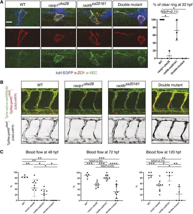
Fig. 7.

Phenotypic comparison of radil-b single and of rasip1/radil-b double mutants suggests partially overlapping functions during vascular morphogenesis. (A) Immunofluorescence analysis of ZO-1 and VE-cadherin distribution in Tg(kdrl:EGFP)s843 at 32 hpf. The graph shows the percentage of DLAVs exhibiting a clear ring at 32 hpf. Number of embryos and junctions analyzed at 32 hpf: WT (5, 15), rasip1ubs28 (3, 11), radil-bsa20161 (3, 6) and double mutant (2, 3). (B) Live images at 48 hpf using Tg(ve-cad:ve-cadVENUS); Tg(fliep:gal4ff)ubs3; (UAS:mRFP) reporter lines. Scale bars: 20 µm. (C) Quantification of blood flow defects in ISVs at 48, 72 and 120 hpf in rasip1ubs28, radil-bsa20161 and in double mutants. radil-bsa20161 mutants show only transient defects in blood flow at 48 hpf. rasip1ubs28; radil-bsa20161 double mutants show a strongly enhanced phenotype. Number of embryos and ISVs analyzed at 48, 72 and 120 hpf, respectively: WT (4, 44; 5, 71; 6, 87), rasip1ubs28 (12, 176; 8, 119; 10, 118), radil-bsa20161 (9, 105; 8, 78; 5, 41) and double mutant (5, 56; 10, 102; 6, 55). Analyzed by unpaired two-tailed Mann–Whitney test (A,C) (NS, no significance; *P<0.1, **P<0.01, ***P<0.001, ****P<0.0001); error bars indicate s.d.

Figure Data
Acknowledgments
This image is the copyrighted work of the attributed author or publisher, and ZFIN has permission only to display this image to its users. Additional permissions should be obtained from the applicable author or publisher of the image. Full text @ Development